








 Work time
Diagnostics: Mon - Sun, 6:00 - 23:00
Polyclinic: Mon - Sun, 8:00 - 20:00

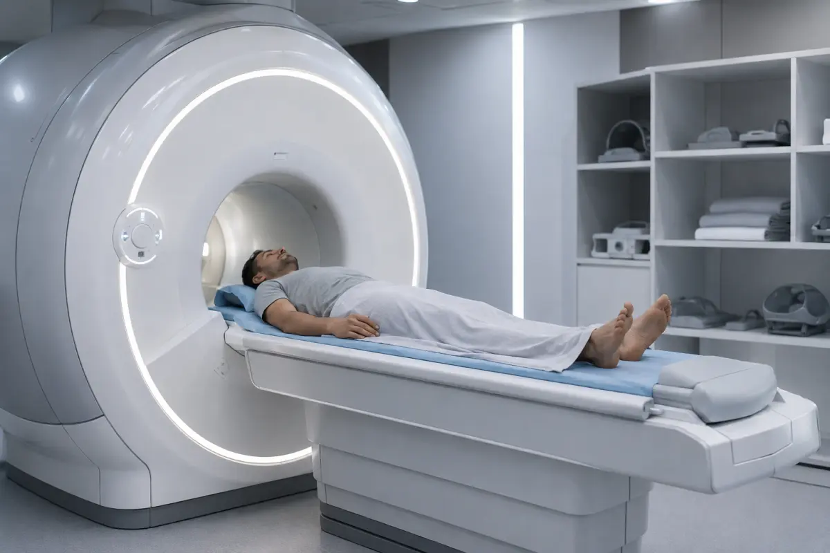
Rectal MRI is a magnetic resonance imaging examination that shows the rectal wall layer by layer, along with the anal canal, surrounding fat tissue, lymph nodes and pelvic floor muscles. It is most often prescribed to assess the spread of a rectal tumor, to locate the course of a fistula in paraproctitis and to monitor the condition after treatment.
At HBMedical medical center, a rectal MRI costs 5400 UAH without contrast and 7800 UAH with contrast. The examination is performed on a Siemens Magnetom Sempra 1.5 Tesla scanner at: Kyiv, 3 Oleksandra Dovzhenka St. This is a 2-minute walk from «Shuliavska» metro station. The diagnostic department is open every day from 6:00 to 23:00.
Rectal MRI Price
The final price depends on several things:
- whether intravenous contrast is needed;
- whether additional areas need to be examined (for example, abdominal organs);
- whether sedation is needed.
A comprehensive MRI of the abdominal and pelvic organs with a single contrast injection costs 9400 UAH.
An autumn promotion on MRI is running, depending on the time of the examination: −25% from 5:30 to 7:00, −15% from 7:00 to 8:00 and −20% from 21:00 to 23:30. Please confirm the terms of the promotion and current prices with the call center when booking.
What a Rectal MRI Shows
MRI distinguishes soft tissues well. That is why the doctor can see not only the rectum itself but also everything surrounding it. The examination makes it possible to assess:
- the thickness and structure of the rectal wall, layer by layer;
- the size of a mass and the depth of its growth into the wall and surrounding tissues;
- the distance from the tumor to the anal canal and sphincters, which is important for surgical planning;
- the condition of the mesorectal fat and the fascia that bounds it;
- pelvic lymph nodes;
- fistulas, abscesses and inflammatory infiltrates, as well as the course of a fistula relative to the sphincter muscles;
- narrowing of the bowel lumen;
- postoperative and scar changes, and signs of recurrence after treatment.
MRI has its limits. The method does not assess the mucosa as thoroughly as colonoscopy. It also does not allow a biopsy to be taken. Small polyps are detected more reliably by endoscopic examination, and the final nature of a mass is determined by histology.
When a Rectal MRI Is Prescribed
A referral is usually issued by a proctologist, oncologist, surgeon or gastroenterologist. MRI often becomes a clarifying step after an examination, colonoscopy or ultrasound.
Typical clinical situations:
- a confirmed or suspected rectal tumor, when the stage and extent of the process need to be determined;
- paraproctitis and rectal fistulas, especially complex or recurrent ones;
- pain in the rectal or perineal area whose cause could not be established by other methods;
- blood in the stool, a feeling of incomplete evacuation, a change in stool pattern — if the doctor considers MRI appropriate;
- surgical planning;
- assessing the result of chemoradiotherapy or surgical treatment;
- dynamic follow-up after treatment.
Symptoms alone do not mean that an MRI is what you need. The method and the sequence of examinations are chosen by the doctor.
Rectal MRI with or without Contrast
In an MRI with contrast, a gadolinium-based agent is injected intravenously. It shows how tissues take up the contrast. This helps to delineate a mass more clearly, assess an inflammatory process or abscess and distinguish scar tissue from active tissue after treatment.
Contrast is not always needed. When assessing a rectal tumor, the main information comes from special high-resolution non-contrast sequences. Therefore, for some clinical questions, an examination without contrast is sufficient.
Follow the referral. If the doctor specified «with contrast», do not choose the non-contrast option on your own because of the price difference: some of the necessary information may be lost.
Before contrast is given, tell staff about kidney disease and any previous reactions to contrast agents. If there are risk factors, you may be asked for a blood test for creatinine.
Rectal MRI, Colonoscopy, CT or Ultrasound
| Method | What it assesses best | Limitations |
| Rectal MRI | The bowel wall layer by layer, tumor spread into tissues, fistulas, pelvic lymph nodes | Cannot see the mucosa from the inside, does not allow a biopsy |
| Colonoscopy | The mucosa, polyps, allows a biopsy to be taken | Does not show what is happening beyond the bowel wall |
| CT | Assessment of distant organs, complications, acute conditions | Distinguishes bowel wall layers and pelvic soft tissues less well, uses X-ray radiation |
| Ultrasound | A quick initial assessment of pelvic organs | Limited depth and detail for the rectum |
These methods do not replace but complement each other. At HBMedical, you can have both an MRI and an endoscopic examination and colonoscopy at one center. This is convenient when the doctor has prescribed both tests.
Preparing for a Rectal MRI
The goal of preparation is to reduce the amount of gas and contents in the bowel. These are what most often reduce image quality. The administrator gives exact instructions when you book, because they depend on the examination protocol.
Preparation usually includes:
- a referral from your doctor and previous results: colonoscopy, histology, CT, MRI, discharge summaries after surgeries or treatment;
- a light diet for 1–2 days without foods that increase gas formation: legumes, raw vegetables, black bread, carbonated drinks;
- not eating for several hours before the procedure;
- emptying the rectum, if this is provided for in the center’s instructions;
- rules about bladder filling, which the administrator will tell you;
- a creatinine test before contrast, if one has been prescribed.
Do not take laxatives or antispasmodics or use an enema on your own without a recommendation from the medical center. Do not stop taking regular medications without consulting your doctor.
How the Examination Is Performed
- You fill in a safety questionnaire and leave metal and electronic items outside the room.
- You lie down on the scanner table, and a receiver coil is placed over the pelvic area.
- The table moves into the scanner. During scanning, you need to lie still and follow the instructions of the MRI technologist, with whom you have contact throughout the procedure.
- If contrast is prescribed, the agent is injected into a vein, after which additional series of images are acquired.
The scanning itself is painless. The machine is loud, so patients are provided with hearing protection. An MRI procedure usually takes from 20 to 40 minutes, and longer with contrast. The scanner is designed for patients weighing up to 220 kg.
Results and Report
A radiologist analyzes the images and prepares a report. You can receive it in 5–7 hours, either in person or through your patient account on the website. The images are saved to a USB flash drive so that you can show them to your doctor.
If a rectal MRI has already been done at another center, HBMedical radiologists can prepare a report based on the existing images. Such a report can be used as a second opinion before surgery or a change of treatment. If needed, the report can be issued in English, for example for a consultation abroad.
Contraindications and Limitations
Be sure to tell staff if you have:
- a pacemaker, defibrillator, neurostimulator, cochlear implant or insulin pump;
- metal devices, vascular clips or fragments in the body;
- pregnancy, especially in the first trimester;
- severe kidney disease, if contrast is planned;
- pronounced claustrophobia or a condition that makes it difficult to lie still.
Having an implant does not always rule out MRI. It all depends on its model and MR compatibility, so bring the documents for the device. If you are afraid of enclosed spaces, the examination can be performed under sedation.
How Rectal MRI Differs from Pelvic MRI
Rectal MRI is a targeted examination. The images are acquired in a way that shows the bowel itself, the anal canal, the sphincters and the pararectal fat in as much detail as possible.
Pelvic MRI covers several organs at once. In men, these are the prostate, bladder and rectum; in women, the uterus, ovaries and bladder. If your referral refers specifically to the pelvis, see the page MRI of male pelvic organs.
When booking, name the examination exactly as the doctor specified it. Do not replace one examination with another on your own.
Rectal MRI in Shuliavka: What to Know about the Center
- Siemens Magnetom Sempra 1.5 Tesla scanner, closed-bore type, manufactured in 2021.
- Diagnostics are available every day from 6:00 to 23:00, so you can schedule the examination before or after work.
- Address: 3 Oleksandra Dovzhenka St., a 2-minute walk from «Shuliavska» metro station. The center is convenient for residents of Shuliavka, KPI, Lukianivka, Dorohozhychi, Nyvky, and the Shevchenkivskyi, Solomianskyi and Sviatoshynskyi districts.
- There is parking near the clinic.
- For people with special needs, lifts, wide corridors and accessible restrooms are provided. Medical transfer across Kyiv is available.
- The polyclinic, laboratory, endoscopy and surgery departments work at one center. After the MRI, you can get a consultation with a specialist without traveling to other facilities.
- The center has its own independent power supply: patients are seen regardless of whether there is electricity in the city.
For a description of all examination areas and the general price list, see the page MRI diagnostics at HBMedical.
How to Book a Rectal MRI
Call +38 (099) 344-30-00 or submit a request through the form on the website, and a consultant will call you back to confirm.
When booking, tell staff:
- the exact name of the examination from the referral and whether contrast is needed;
- whether you have implants or metal devices;
- whether you have kidney disease or claustrophobia;
- whether you have previous MRI, CT, colonoscopy or histology results.
The administrator will select a time and provide preparation instructions.
Frequently Asked Questions
How much does a rectal MRI cost at HBMedical?
Without contrast — 5400 UAH, with contrast — 7800 UAH. The price includes the radiologist’s report and a USB flash drive with the examination recording.
What does a rectal MRI show?
The structure of the bowel wall, the size and spread of masses, the condition of the pararectal fat and lymph nodes, fistulas, abscesses, narrowing, postoperative changes and signs of recurrence.
How do I prepare for a rectal MRI?
You are usually asked to follow a light diet without gas-forming foods for 1–2 days, not to eat for several hours before the procedure and, if necessary, to empty the rectum. You will get the exact rules when you book.
Can I eat before the examination?
You are usually asked not to eat for several hours before the procedure, especially if contrast is planned. Confirm the exact interval when booking.
Is contrast mandatory?
No. The need for contrast is determined by the doctor depending on the clinical question. For some examinations, a non-contrast protocol is sufficient.
Can a rectal MRI replace a colonoscopy?
No. Colonoscopy assesses the mucosa and allows a biopsy to be taken, while MRI shows the bowel wall and the tissues around it. Doctors often prescribe both examinations.
How long does a rectal MRI take?
The scan usually takes 20–40 minutes. With contrast, the procedure takes longer because a catheter has to be placed and additional series of images are needed.
When will the report be ready?
Within 5–7 hours. You can pick it up in person or receive it through your patient account on the website.
Is a rectal MRI painful?
The scanning itself is painless. When contrast is given, you only feel the prick of placing a catheter in a vein.
What if I am afraid of enclosed spaces?
Tell staff when you book. At HBMedical, MRI under sedation is possible.
Can I have an MRI without a referral?
Please confirm this option when booking. A referral is advisable because it helps to choose the right protocol and decide on contrast.
Can I get a report for an MRI done at another clinic?
Yes. HBMedical radiologists prepare reports for examinations performed at another center. The cost is 2000 UAH.
The information on this page is for reference only and does not replace a doctor’s consultation.






















Мені все сподобалось
Всі профі
Дякую


Велика подяка лікарю Дуброву Є.О. — надзвичайно компетентний і чуйний фахівець, який детально все пояснив, розвіяв тривогу та підтримав морально. Щиро дякую анестезіологу Бєлаш О.М. за високий професіоналізм, чуйність і справжнє людське ставлення — занурення в сон та сам перебіг процедури минули максимально комфортно. Також величезне спасибі медсестрі (на жаль, не дізналася ім'я) за її турботу, доброту та увагу.
Весь персонал вразив своєю чуйністю та теплом. Дякую за ваш труд і турботу про пацієнтів! Однозначно рекомендую.













Дуже ввічливий та приємний персонал клініки. Лікар був уважний, без поспіху пройшовся по всім аспектам здоровʼя. Лабораторія швидко зробила аналізи. В цілому дуже задоволена. Радже звертатися!




Саме до таких фахівців, в разі необхідності, є бажання повернутися!

і закінчуючи отриманим описом і рекомендаціями від лікарів.
























Need specialist help?
Order a callback and our consultant will help you choose a doctor and make an appointment at a convenient time for you!