


![]() Work time
Work time
Diagnostics: Mon - Sun, 6:00 - 23:00
Polyclinic: Mon - Sun, 8:00 - 20:00
A CT scan of joints is a modern computed tomography examination that helps assess the bone structures of a joint, articular surfaces, consequences of injuries, degenerative changes, inflammatory processes, and the condition after surgery or the installation of metal implants in detail. At the HBMedical medical center in Kyiv, you can have a CT scan of one joint, two paired joints, or a separate anatomical area near Shuliavska metro station.
Computed tomography of joints is especially useful when a standard X-ray is not enough, pain does not go away, there is a suspected fracture, fissure, displacement of bone fragments, arthrosis, complications after an injury, or when it is necessary to clarify the condition of the joint before treatment. You can book a CT scan of joints at HBMedical online or by phone.
What is a CT scan of joints?
A CT scan of joints is a layer-by-layer X-ray scan during which the scanner creates detailed images of the examined area. Unlike a standard X-ray, computed tomography makes it possible to see the joint in different planes and better assess complex anatomical structures.
During a CT scan, the doctor can examine bones, articular surfaces, small fractures, fissures, bone growths, deformities, and the consequences of injuries or surgeries in detail. If necessary, 3D reconstruction can be performed to better understand the position of bone structures and plan further treatment.
CT scans of joints are most often prescribed by orthopedic traumatologists, surgeons, rheumatologists, sports medicine doctors, or family doctors when it is necessary to quickly and accurately clarify the cause of pain, restricted movement, or changes in a joint.
Which joints can be examined with CT?
At HBMedical, you can undergo computed tomography of different joints and anatomical areas. The type of examination depends on the complaints, injury, doctor’s referral, and the specific area that needs to be assessed.
Most often, patients book:
- CT scan of the knee joint;
- CT scan of the hip joint;
- CT scan of the hip joint;
- CT scan of the shoulder joint;
- CT scan of the elbow joint;
- CT scan of the wrist joint;
- CT scan of the ankle joint;
- CT scan of the hand;
- CT scan of the foot;
- CT scan of joints after injury;
- CT scan of a joint with a metal implant;
- CT scan of two paired joints for comparison.
If the referral does not specify a particular joint but says “CT scan of an anatomical area,” the administrator will help correctly determine the service based on the doctor’s referral during booking.
What does computed tomography of joints show?
A CT scan of joints helps assess mainly bone structures and changes that may be poorly visible on a standard X-ray. The examination can show:
- fractures, fissures, and microfractures;
- displacement of bone fragments;
- consequences of dislocations and subluxations;
- arthrosis and degenerative changes in the joint;
- bone growths, osteophytes;
- deformation of articular surfaces;
- inflammatory changes in bone tissue;
- signs of osteomyelitis;
- bone cysts or neoplasms;
- developmental abnormalities of the joint;
- condition after surgery;
- position of metal implants;
- complications after injuries or surgical treatment.
A CT scan of joints is not always the main method for assessing ligaments, menisci, tendons, and cartilage, so in some cases the doctor may additionally recommend MRI or ultrasound. But if it is necessary to see bones, articular surfaces, fractures, or the consequences of an injury in detail, CT is one of the most informative methods.
When is a CT scan of a joint needed?
Computed tomography of a joint may be prescribed after an injury, in cases of chronic pain, restricted mobility, or before surgery. CT is also often used when X-ray results are not informative enough or when symptoms do not match previous images.
A CT scan of joints is recommended for the following complaints and situations:
- joint pain after a fall, impact, or sports injury;
- suspected fracture, fissure, dislocation, or subluxation;
- swelling, deformity, or sudden restriction of movement;
- long-term pain without a clear cause;
- crunching, instability, or a feeling of joint locking;
- suspected arthrosis or pronounced degenerative changes;
- preparation for joint surgery;
- follow-up after surgical treatment;
- checking the position of plates, screws, endoprostheses, or other metal implants;
- clarification of X-ray, ultrasound, or MRI results;
- suspected tumor or inflammatory changes in bone tissue.
If joint pain appeared after an injury, the joint is deformed, the limb cannot bear weight, or the pain suddenly worsens, diagnostics should not be postponed. In such cases, you should see a doctor as soon as possible.
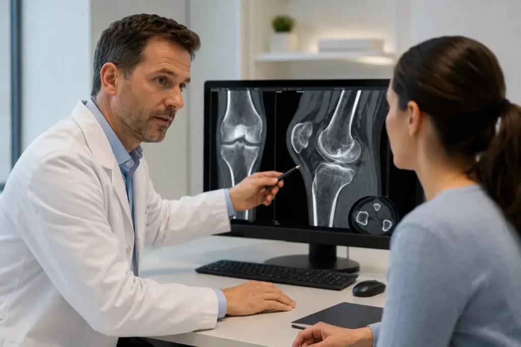
CT scan of the knee joint
A CT scan of the knee joint is often prescribed after injuries, falls, impacts, sports activities, or when a complex fracture is suspected. The knee has a complex structure and is constantly under load during walking, running, squatting, and climbing stairs.
Computed tomography of the knee helps assess the bones that form the joint, articular surfaces, consequences of fractures, fissures, displacement of fragments, arthritic changes, and the condition after surgery. A CT scan of the knee joint can also be useful before surgical treatment when the doctor needs to understand the anatomy of the injury precisely.
CT scan of the hip joint
A CT scan of the hip joint is prescribed for pain in the groin, thigh, or pelvis, after falls, when a femoral neck fracture, arthrosis, aseptic necrosis, or complications after endoprosthetic replacement are suspected.
This examination is especially important for older patients after injuries, when an X-ray does not provide a complete answer, as well as for patients with installed metal structures. CT allows the doctor to assess bone structures, the position of implants or prostheses, the degree of changes, and possible complications.
CT scan of the shoulder joint
A CT scan of the shoulder joint may be needed after a fall on the arm or shoulder, when a fracture, dislocation, damage to the glenoid cavity, bone growths, or post-traumatic changes are suspected. The shoulder joint has a wide range of motion, so it is often injured during sports, physical work, or everyday falls.
A shoulder CT scan helps the doctor assess bone structures, consequences of injuries, the shape of articular surfaces, and possible causes of restricted movement.
CT scan of the elbow joint
A CT scan of the elbow joint is prescribed for arm injuries, falls on the elbow, suspected intra-articular fracture, restricted flexion or extension, chronic pain, and consequences of dislocation or surgery.
The elbow joint has complex anatomy, so small fractures or displacements are not always clearly visible on a standard X-ray. CT helps clarify the nature of the injury and choose the right treatment strategy.
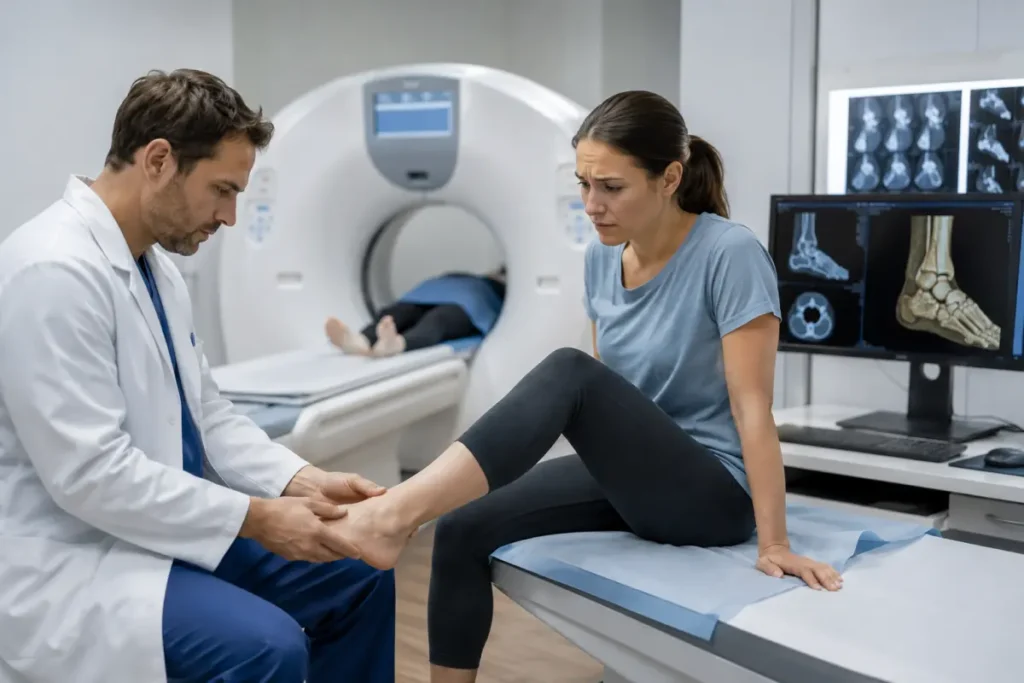
CT scan of the ankle joint, foot, and hand
A CT scan of the ankle joint is often needed after twisting the foot, a fall, a sports injury, or when a fracture of the malleoli, talus, calcaneus, or other foot structures is suspected. A CT scan of the foot can be useful in cases of complex fractures, deformities, pain after injury, or before surgery.
A CT scan of the hand and wrist joint is prescribed for hand injuries, suspected fractures of small bones, chronic pain, restricted movement, or follow-up after treatment.
CT scan of joints with contrast
In most cases, CT scans of joints are performed without contrast, especially when it is necessary to assess a fracture, arthrosis, bone changes, the position of a metal implant, or the consequences of an injury. Contrast may be needed less often — for example, if the doctor needs to clarify the nature of a soft-tissue process, inflammatory changes, a neoplasm, or vascular features in the examined area.
You should not choose CT with contrast on your own “for better accuracy.” Contrast enhancement must be justified. If the referral indicates a CT scan with contrast, before the procedure you should inform the medical staff about allergies, chronic diseases, kidney condition, and the medications you take.
Price of CT scans of joints at HBMedical
HBMedical has a clear price list for computed tomography of joints. The cost depends on the number of joints, the examination area, the use of contrast, and the complexity of diagnostics.
| Service | Without contrast | With contrast |
| CT scan of one joint | UAH 1850 | UAH 3000 |
| CT scan of two paired joints | UAH 2600 | UAH 4050 |
| CT scan of one anatomical area | UAH 2500 | UAH 3400 |
| CT scan of an upper or lower limb area with a metal implant | UAH 3950 | not performed |
The price of the examination includes the doctor’s report and a flash drive. It is better to confirm the current price of a CT scan of joints in Kyiv during booking, since the final type of service depends on the doctor’s referral and the examination area.
How is a CT scan of a joint performed?
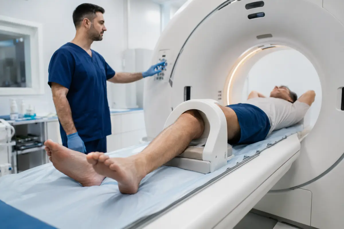
The CT scan procedure is quick and painless. The patient arrives at the medical center at the appointed time, completes registration, and provides the doctor’s referral and previous examinations, if available. Before scanning, metal objects should be removed from the examination area whenever possible.
During the examination, the patient lies on the CT scanner table. The joint area is positioned so that the scanner can perform an accurate scan. It is important to remain still because movement can reduce image quality. After scanning, the radiologist analyzes the images and prepares a report.
If the CT scan is performed with contrast, before the procedure the medical staff additionally checks contraindications, creatinine test results, allergic reactions, and places an intravenous catheter for contrast agent administration.
How to prepare for computed tomography of joints?
No special preparation is usually required for a CT scan of a joint without contrast. It is enough to arrive at the appointed time and bring the doctor’s referral, documents, and previous examination results.
Before the CT scan, it is advisable to bring:
- a doctor’s referral, if available;
- previous X-ray images, CT, MRI, or ultrasound results;
- discharge summaries after injuries, surgeries, or treatment;
- documents related to installed metal implants or endoprostheses, if available;
- comfortable clothing without metal elements in the examination area.
If a CT scan with contrast is planned, a blood creatinine test may be required. You should also report allergies to iodine or contrast agents, kidney disease, thyroid disease, diabetes, pregnancy, or breastfeeding.
Can a CT scan of a joint be done with metal implants?
Yes, CT is often used to assess joints and bones after surgery when there are plates, screws, pins, endoprostheses, or other metal structures in the examined area. Such an examination helps assess the position of the structure, the condition of bone tissue, signs of displacement, complications, or the healing process.
HBMedical has a separate service: CT scan of an upper or lower limb area with a metal implant. If you have metal in the examination area, be sure to mention this during booking so that the administrator can select the correct service.
CT or MRI of joints: which is better?
CT and MRI of joints are not complete equivalents. Each method has its own strengths.
CT is better suited for assessing bones, fractures, fissures, articular surfaces, bone growths, consequences of injuries, metal implants, and preparation for surgery. MRI is more often chosen when it is necessary to assess soft tissues in detail: ligaments, tendons, menisci, cartilage, muscles, inflammatory changes, or edema.
If a fracture is suspected after an injury or bone damage needs to be clarified, the doctor may prescribe CT. If the main concern involves ligaments, menisci, or cartilage, MRI may be needed. The correct diagnostic method is best chosen based on a doctor’s recommendation.
CT scan of joints after injury
After joint injuries, CT helps quickly clarify the nature of the damage. This is especially important in complex fractures, intra-articular injuries, suspected displacement of fragments, or when pain persists but X-ray does not show obvious changes.
CT after an injury may be needed after a fall, traffic accident, sports injury, impact, ankle sprain, fall on the arm, pain after physical exertion, or inability to move the limb normally. The more precisely the doctor sees the injury, the easier it is to choose the right treatment and reduce the risk of complications.
Who may have contraindications to CT scans of joints?
CT uses X-ray radiation, so the examination is not performed without need and is prescribed based on medical indications. The main limitation is pregnancy. Difficulties may also arise if the patient cannot remain still due to severe pain or another condition.
CT with contrast has additional restrictions:
- allergy to iodine-containing contrast;
- severe kidney failure;
- certain thyroid diseases;
- decompensated diabetes mellitus;
- severe reactions to contrast in the past;
- pregnancy or breastfeeding.
If you have chronic diseases or previous reactions to contrast, report this during booking.
Why choose HBMedical near Shuliavka for a CT scan of joints?
HBMedical is a modern clinical and diagnostic center in Kyiv where you can undergo a CT scan of joints in a convenient location near Shuliavska metro station. The center is suitable for patients who need to quickly clarify the cause of pain, check a joint after an injury, prepare for a consultation with an orthopedic traumatologist, or monitor their condition after surgery.
Advantages of HBMedical:
- convenient location near Shuliavska metro station;
- address: Kyiv, 3 O. Dovzhenka St.;
- modern Siemens CT scanner;
- CT scan of one joint, two paired joints, or an anatomical area;
- possibility of examining an area with a metal implant;
- appointment online or by phone;
- clear CT scan prices;
- diagnostics daily from 6:00 to 23:00;
- doctor’s report and flash drive included in the price;
- possibility to receive an additional consultation or examination in one medical center.
The location near Shuliavka is convenient for patients from Shevchenkivskyi, Solomianskyi, Sviatoshynskyi districts, KPI, Nyvky, Lukianivka, Dorohozhychi, and other areas of Kyiv.
How to book a CT scan of joints in Kyiv?
To book a CT scan of joints at HBMedical, leave a request on the website or call the medical center. During booking, it is advisable to specify which joint needs to be examined: knee, hip, shoulder, elbow, ankle, wrist, hand, foot, or another area.
If you have a referral, tell the administrator its wording. This will help select the correct service: CT scan of one joint, CT scan of two paired joints, CT scan of an anatomical area, or CT scan of an area with a metal implant.
HBMedical — CT scan of joints in Kyiv
Address: Kyiv, 3 O. Dovzhenka St., Shuliavska metro station
Service: computed tomography of joints
Price: from UAH 1850
Appointment: online or by phone
Frequently asked questions about CT scans of joints
How much does a CT scan of a joint cost in Kyiv?
At HBMedical, a CT scan of one joint costs from UAH 1850 without contrast. A CT scan of one joint with contrast costs UAH 3000. A CT scan of two paired joints costs from UAH 2600. It is better to confirm the current price during booking.
Where can I have a CT scan of joints near Shuliavka?
You can undergo a CT scan of joints at the HBMedical medical center in Kyiv at 3 O. Dovzhenka St., near Shuliavska metro station.
Which joints can be examined with CT?
Most often, CT scans are performed for the knee, hip, shoulder, elbow, ankle, and wrist joints, as well as CT scans of the hand, foot, or a separate anatomical area.
Is preparation needed for a CT scan of a joint?
No special preparation is usually required for a CT scan of a joint without contrast. You should bring the doctor’s referral and previous examinations, if available. For CT with contrast, a blood creatinine test may be required.
Is a CT scan of joints painful?
No, the procedure is painless. The patient simply lies still during scanning. Discomfort may occur only because it is necessary to maintain a certain position, especially if the joint hurts after an injury.
How long does a CT scan of a joint take?
The scan itself is quick, but you should also allow time for registration, preparation for the procedure, and waiting for the results. It is better to confirm the approximate duration during booking.
Are CT scans of joints performed with or without contrast?
In most cases, CT scans of joints are performed without contrast. Contrast may be needed in certain clinical situations, for example, when a neoplasm, a complex inflammatory process, or soft-tissue changes need to be clarified.
Can a CT scan of a joint be done with a metal implant?
Yes, CT is often used for follow-up after surgery and when metal implants are present. HBMedical has a separate service for CT scans of an upper or lower limb area with a metal implant.
What is better after an injury: X-ray or CT?
X-ray is often the first stage of diagnostics. If it does not provide enough information or there is a suspicion of a complex fracture, fissure, intra-articular injury, or displacement of fragments, the doctor may prescribe CT.
Can I have a CT scan of joints without a referral?
It is advisable to have a doctor’s referral to correctly determine the area and purpose of the examination. If there is no referral, it is worth consulting an orthopedic traumatologist or clarifying during booking which diagnostic option may be needed.


Саме до таких фахівців, в разі необхідності, є бажання повернутися!


Need specialist help?
Order a callback and our consultant will help you choose a doctor and make an appointment at a convenient time for you!