








 Work time
Diagnostics: Mon - Sun, 6:00 - 23:00
Polyclinic: Mon - Sun, 8:00 - 20:00

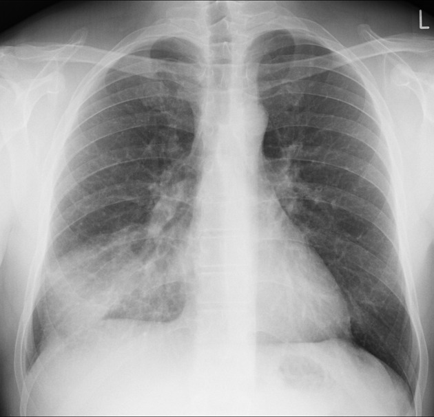
Radiography is a fast and informative diagnostic imaging method used to assess the condition of bones, joints, the spine, chest organs, and other anatomical structures. It can help detect fractures, deformities, degenerative changes, abnormal bone alignment, and other pathological conditions.
The images below are examples of X-ray examinations. The final interpretation of the results is performed by a radiologist, taking into account the patient’s symptoms, medical history, and overall clinical presentation.
Pelvic Radiography

Radiological description: signs of deforming osteoarthritis of the right hip joint, stage II, and the left hip joint, stage III–IV, according to the Kellgren–Lawrence classification. Sequelae of a fracture of the left acetabulum.
Cervical Spine Radiography with Functional Views


Radiological description: no radiographic signs of cervical spine instability were detected. Deforming spondylosis at the C4–C7 levels. Moderate degenerative changes in the cervical spine.
Standing Long-Leg Radiography of Both Lower Limbs
Standing long-leg radiography helps assess the alignment and length of the lower limbs, joint positioning, and the presence of deformities.

Radiological description: signs of varus deformity of the left lower limb. Coxa valga of both hip joints. Stage II osteoarthritis of both hip joints and stage I osteoarthritis of both knee joints.
Shoulder Joint Radiography

Radiological description: signs of stage II osteoarthritis of the shoulder joints according to the Kellgren–Lawrence classification. Cystic remodelling of the left humeral head. The detected changes may correspond to calcific tendinitis in the projection of the right supraspinatus tendon.
Signs of osteoarthritis of the right acromioclavicular joint and asymmetric widening of the left acromioclavicular joint are also present.
Lumbar Spine Radiography with Functional Views


Radiological description: signs of degenerative disc disease of the lumbar spine.
Full-Spine Radiography
Full-spine radiography makes it possible to assess spinal alignment, the degree of curvature, vertebral rotation, and coronal and sagittal balance.

Radiological description: signs of grade II S-shaped scoliosis of the thoracolumbar spine. Grade I thoracic hyperkyphosis. Grade II rotation of the lumbar vertebral bodies according to Raimondi. Abnormal coronal and sagittal balance.
Spinal Radiography after Transpedicular Instrumentation

Radiological description: status after bilateral transpedicular instrumentation at the Th4–L1 levels. Signs of grade I S-shaped scoliosis of the thoracolumbar spine. Abnormal coronal and sagittal balance. Straightening of the physiological lordosis.
Lumbar Spine Radiography after Fixation

Radiological description: signs of grade I L5 spondylolisthesis according to the Meyerding classification. Status after transpedicular fixation at the L4–S1 levels. Signs of degenerative disc disease of the lumbar spine.
Left Hand Radiography

Radiological description: no radiographic signs of pathological changes in the left hand were detected.
Left Ankle Radiography

Radiological description: signs of a fibular fracture at the level of the distal tibiofibular syndesmosis without significant displacement. Signs of an impacted fracture of the talar head with minimal displacement.
Important: a radiological report is not a final clinical diagnosis. The results of the examination should be reviewed with a doctor, who will take into account the patient’s symptoms, medical history, and the results of other examinations.










Мені все сподобалось
Всі профі
Дякую


Велика подяка лікарю Дуброву Є.О. — надзвичайно компетентний і чуйний фахівець, який детально все пояснив, розвіяв тривогу та підтримав морально. Щиро дякую анестезіологу Бєлаш О.М. за високий професіоналізм, чуйність і справжнє людське ставлення — занурення в сон та сам перебіг процедури минули максимально комфортно. Також величезне спасибі медсестрі (на жаль, не дізналася ім'я) за її турботу, доброту та увагу.
Весь персонал вразив своєю чуйністю та теплом. Дякую за ваш труд і турботу про пацієнтів! Однозначно рекомендую.














Дуже ввічливий та приємний персонал клініки. Лікар був уважний, без поспіху пройшовся по всім аспектам здоровʼя. Лабораторія швидко зробила аналізи. В цілому дуже задоволена. Радже звертатися!




Саме до таких фахівців, в разі необхідності, є бажання повернутися!

і закінчуючи отриманим описом і рекомендаціями від лікарів.























Need specialist help?
Order a callback and our consultant will help you choose a doctor and make an appointment at a convenient time for you!