


![]() Work time
Work time
Diagnostics: Mon - Sun, 6:00 - 23:00
Polyclinic: Mon - Sun, 8:00 - 20:00
MRI of the hip joints — is a magnetic resonance imaging examination of the hip area that helps assess bone structures, articular cartilage, the femoral head, acetabulum, tendons, muscles, ligaments, labrum and periarticular soft tissues in detail. At the HBMedical medical center in Kyiv, you can undergo MRI of the hip joints near Shuliavska metro station — by prior online or phone appointment.
The examination may be needed for pain in the hip joint, groin, buttock or upper thigh, after an injury, in cases of suspected inflammation, osteoarthritis, avascular necrosis, labral tear, bursitis, tendinitis or other changes that need to be clarified after an examination, ultrasound, X-ray or CT.
What is MRI of the hip joints?
MRI of the hip joints — is a diagnostic method that uses a magnetic field and radio waves to create detailed images of the hip area. Unlike X-ray or CT, MRI does not use ionizing radiation and is better suited for assessing soft tissues, cartilage, tendons, ligaments, bone marrow and periarticular structures.
The hip joint — is one of the largest and most weight-bearing joints in the body. It is involved in walking, running, climbing stairs, bending and maintaining balance. Therefore, pain in this area can significantly limit mobility and quality of life. MRI helps the doctor see not only obvious bone changes but also early or hidden pathological processes that are not always visible on X-ray.
Patients often search for this service as “hip joint MRI”, “MRI of the hip joints Kyiv” or “hip joint MRI price”. In the HBMedical price list, the service is listed as MRI of the hip joints, meaning an examination of the hip area according to the appropriate protocol.
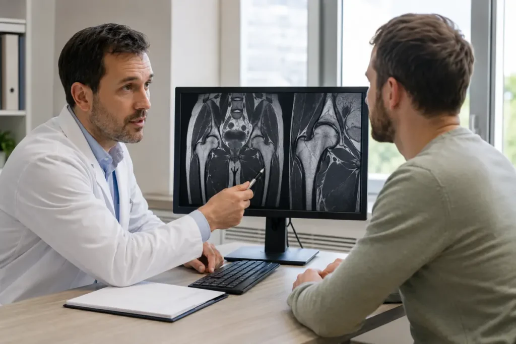
What does an MRI of the hip joint show?
MRI of the hip joint shows structures that are difficult to fully assess on a standard X-ray. The method allows examination not only of the bone surfaces but also cartilage, soft tissues, tendons, ligaments, the labrum, bone marrow and surrounding structures.
MRI of the hip joints may show:
- signs of hip osteoarthritis;
- damage or thinning of articular cartilage;
- inflammatory changes in the joint;
- synovitis, fluid in the joint;
- bursitis in the hip joint area;
- tendinitis or tendon injury;
- sprain or ligament injury;
- labral tear;
- avascular necrosis of the femoral head;
- stress or occult fractures;
- bone marrow edema;
- consequences of injuries;
- postoperative changes;
- enlarged lymph nodes or soft tissue changes in the examination area;
- signs of an infectious or tumor process if clinically indicated.
MRI is not a diagnosis in itself. After the examination, the results should be shown to the doctor who referred you for diagnostics: an orthopedist, traumatologist, rheumatologist, neurologist, surgeon, sports physician or another relevant specialist.
When is MRI of the hip joints prescribed?
MRI of the hip joints may be prescribed for pain, stiffness, limited movement, suspected inflammatory or degenerative process, after an injury or when other diagnostic methods have not provided enough information.
The examination may be recommended if there is:
- pain in the hip joint;
- pain in the groin, buttock or upper thigh;
- pain while walking or climbing stairs;
- limping;
- joint stiffness;
- limited abduction or flexion of the leg;
- pain after a fall or sports injury;
- suspected labral tear;
- suspected avascular necrosis of the femoral head;
- suspected occult or stress fracture;
- suspected bursitis or tendinitis;
- osteoarthritis or suspected degenerative changes;
- inflammatory rheumatologic diseases;
- follow-up after treatment or surgery;
- the need to clarify ultrasound, X-ray or CT results.
If hip joint pain appeared after a serious injury and is accompanied by inability to put weight on the leg, severe limitation of movement, fever, significant swelling or a sudden deterioration in condition, emergency medical care should be sought.
MRI of the hip joints for groin pain
Groin pain is not always related only to muscles or ligaments. Sometimes the cause may be changes in the hip joint, labral injury, tendon inflammation, bursitis, osteoarthritis, overload or problems in the area where muscles attach.
MRI may be useful if groin pain:
- worsens while walking;
- occurs when rotating the leg;
- radiates to the thigh or buttock;
- appeared after training, a fall or a sudden movement;
- does not resolve after treatment;
- is accompanied by limited movement in the hip joint;
- requires clarification after ultrasound or X-ray.
If the doctor suspects ARS syndrome or a problem in the groin area, an extended examination may be prescribed — MRI of the hip joints and groin area for ARS syndrome.
MRI of the hip joint after injury
After injuries to the hip area, MRI helps assess not only bones but also soft tissues. This is important after a fall, sports injury, impact, sudden leg rotation or overload.
MRI of the hip joint after injury may help detect:
- occult fractures that are not visible on X-ray;
- bone marrow edema;
- cartilage damage;
- labral tear;
- ligament sprain;
- tendon injury;
- bursitis or inflammatory changes;
- consequences of old injuries.
If pain does not go away after an injury, limping appears or it becomes difficult to put weight on the leg, it is worth consulting a traumatologist or orthopedist and undergoing an examination as prescribed by the doctor.
MRI of the hip joint for osteoarthritis
Hip osteoarthritis — is a degenerative joint disease in which cartilage, bone surfaces and periarticular structures gradually change. In the early stages, symptoms may be mild, but over time pain, stiffness, limited movement and discomfort while walking may appear.
MRI can help the doctor assess:
- the condition of articular cartilage;
- the presence of inflammation;
- fluid in the joint;
- the condition of bone marrow;
- associated changes in tendons and soft tissues;
- complications or atypical causes of pain.
In pronounced osteoarthritis, the doctor may also use X-ray or CT, especially when bone deformities need to be assessed. However, MRI provides more information about soft tissue and intra-articular changes.
MRI of the hip joints with and without contrast
In most cases, MRI of the hip joints is performed without contrast. This is often sufficient to assess cartilage, bone marrow, tendons, ligaments, the synovial membrane, bursae, inflammatory changes, consequences of injury or degenerative processes.
MRI of the hip joints with contrast is not needed for everyone. Contrast enhancement is prescribed when the doctor needs to clarify a tumor process, active inflammation, postoperative changes, infectious involvement or a complex clinical case where information without contrast may be insufficient.
You should not choose contrast on your own “for accuracy”. Contrast must be justified by the medical task. If the referral states MRI with contrast, you should report kidney diseases, allergic reactions, pregnancy, breastfeeding and medications you take when booking.
MRI of the hip joints price at HBMedical
At HBMedical, the price of MRI of the hip joints depends on whether contrast enhancement is required, whether there is an endoprosthesis or another metal structure, and whether the groin area also needs to be assessed.
| Service | Without contrast | With contrast |
| MRI of the hip joints | UAH 3800 | UAH 6500 |
| MRI of the hip joints and groin area for ARS syndrome | UAH 6400 | UAH 8500 |
| MRI of both hip joints with an endoprosthesis or another metal structure | UAH 5300 | not performed |
The examination price includes the doctor’s report and a flash drive. It is better to clarify the current price of hip joint MRI when booking, because the final service depends on the doctor’s referral, the need for contrast enhancement and the presence of metal structures.
MRI of the hip joint with an endoprosthesis
If a patient has a hip endoprosthesis or another metal structure, this must be reported when booking. Not all metal structures behave the same way in a magnetic field, and the presence of metal may affect image quality.
HBMedical offers a separate service: MRI of both hip joints with an endoprosthesis or another metal structure. This examination may be needed to assess soft tissues around the endoprosthesis, postoperative changes, pain after surgery or other conditions according to a doctor’s referral.
Before MRI with an endoprosthesis, it is advisable to bring documents about the implant type or a postoperative discharge summary. This will help medical staff assess the possibility and safety of performing the examination.
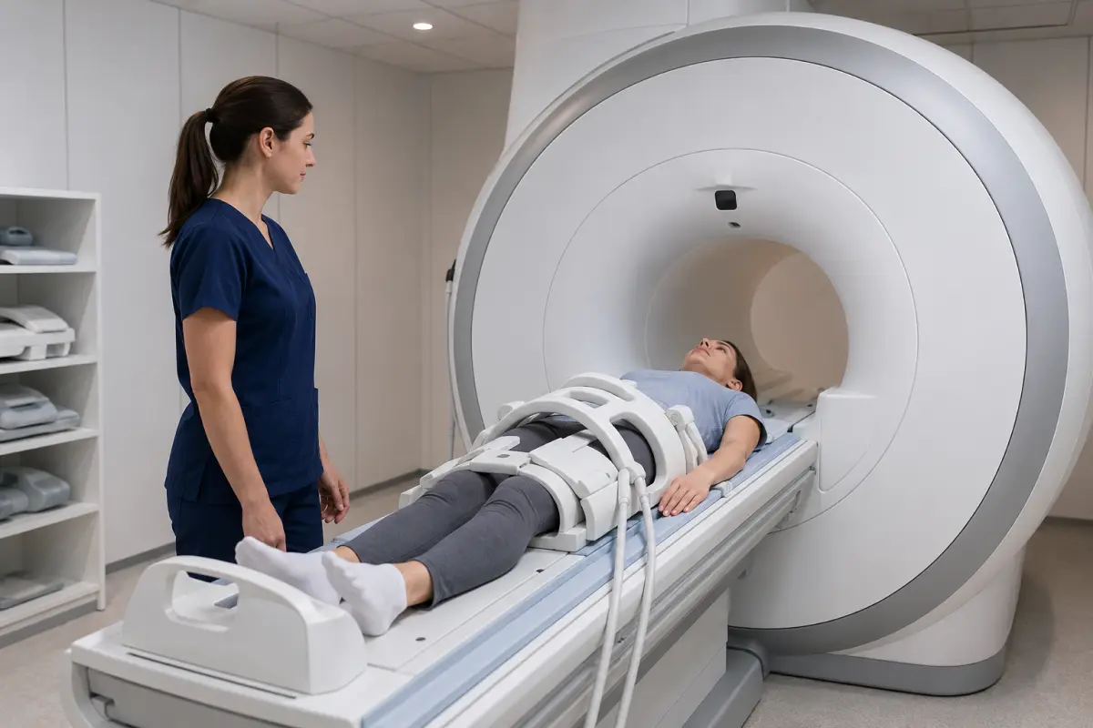
How is MRI of the hip joints performed?
The patient arrives at the medical center at the scheduled time, completes registration and fills out an MRI safety questionnaire. Medical staff check whether there are any contraindications to magnetic resonance imaging, implants or metal objects in the body.
Before the procedure, metal items, electronics, jewelry, watches, bank cards and other items that cannot be brought into the MRI room must be left outside. Then the patient lies down on the scanner table. During scanning, it is important to remain still because movement may reduce image quality.
During operation, the machine makes loud sounds — this is a normal part of MRI. If needed, the patient may be provided with noise-reducing accessories. If the examination is performed with contrast, the contrast agent is administered intravenously through a catheter.
After the procedure, a radiologist analyzes the images and prepares a report. The results should be taken to the doctor who referred you for the examination.
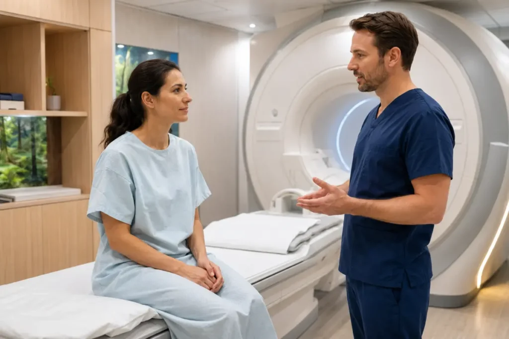
How to prepare for MRI of the hip joint?
For MRI of the hip joints without contrast, no special complex preparation is usually required. The main thing is to inform the staff in advance about implants and metal structures and to bring previous examination results.
Before MRI of the hip joints, it is advisable to:
- bring your doctor’s referral if you have one;
- bring previous ultrasound, X-ray, CT, MRI, discharge summaries or laboratory test results;
- report an endoprosthesis, metal structures, clips, implants or foreign metal bodies;
- report pregnancy or possible pregnancy;
- wear comfortable clothing without metal elements;
- remove jewelry, piercings, watch and metal accessories;
- do not bring a phone, bank cards, keys or electronic devices into the MRI room;
- clarify when booking whether additional preparation is needed in your specific case.
If MRI with contrast is prescribed, kidney function may need to be checked, in particular with a blood creatinine test. You should also report allergic reactions, kidney diseases, pregnancy or breastfeeding.
MRI or CT of the hip joint — which is better?
MRI and CT of the hip joint have different diagnostic capabilities. MRI is better suited for assessing cartilage, soft tissues, tendons, ligaments, the labrum, bone marrow, inflammation, avascular necrosis and hidden traumatic changes.
CT better shows bone structures, complex fractures, bone deformities, the condition after injuries or preparation for certain orthopedic interventions. X-ray is often used as the first method when osteoarthritis or fracture is suspected, but it does not always show soft tissue changes.
It cannot be said that one method is always better. The choice between MRI, CT and X-ray depends on symptoms, the doctor’s referral and the exact question that needs to be answered.
Why have an MRI of the hip joints at HBMedical near Shuliavka?
HBMedical — is a modern medical center in Kyiv where you can undergo MRI of the hip joints on a Siemens Magnetom Sempra 1.5 Tesla scanner. The center is conveniently located near Shuliavska metro station, which is suitable for patients from Shevchenkivskyi, Solomianskyi, Sviatoshynskyi districts, KPI, Nyvky, Lukianivka, Dorohozhychi and other areas of Kyiv.
Advantages of HBMedical:
- convenient location near Shuliavska metro station;
- address: Kyiv, 3 O. Dovzhenka St.;
- modern Siemens Magnetom Sempra 1.5 Tesla MRI scanner;
- MRI of the hip joints with and without contrast;
- possibility of examining the hip joints and groin area for ARS syndrome;
- a separate service for patients with an endoprosthesis or metal structure;
- online or phone booking;
- diagnostics daily from 6:00 to 23:00;
- clear MRI of the hip joints pricing;
- the price includes the doctor’s report and a flash drive;
- possibility to undergo additional examinations or consultations in one medical center.
How to book MRI of the hip joints in Kyiv?
To book MRI of the hip joints at HBMedical, leave a request on the website or call the medical center. When booking, it is advisable to provide the exact wording from the referral: MRI of the hip joints, hip joint MRI with contrast, MRI of the hip joints and groin area for ARS syndrome or MRI with an endoprosthesis.
The administrator will help you choose a convenient time, clarify the current price, explain preparation and advise which documents or previous examination results you should bring with you.
HBMedical — MRI of the hip joints in Kyiv
Address: Kyiv, 3 O. Dovzhenka St., Shuliavska metro station
Services: MRI of the hip joints, MRI of the hip joints with contrast, MRI with an endoprosthesis
Price: from UAH 3800
Booking: online or by phone
Frequently asked questions about MRI of the hip joints
How much does MRI of the hip joints cost in Kyiv?
At HBMedical, MRI of the hip joints costs UAH 3800 without contrast and UAH 6500 with contrast. It is better to clarify the current price when booking.
What is the price of hip joint MRI without contrast?
In the HBMedical price list, the service is listed as MRI of the hip joints. The cost without contrast is UAH 3800. The price includes the doctor’s report and a flash drive.
How much does hip joint MRI with contrast cost?
MRI of the hip joints with contrast at HBMedical costs UAH 6500. Contrast enhancement is performed according to indications or a doctor’s referral.
Where can I have MRI of the hip joints near Shuliavka?
MRI of the hip joints can be performed at the HBMedical medical center in Kyiv at the address: 3 O. Dovzhenka St., near Shuliavska metro station.
What does hip joint MRI show?
Hip joint MRI shows cartilage, bone marrow, the femoral head, acetabulum, tendons, ligaments, muscles, the labrum, fluid in the joint and periarticular soft tissues.
When is hip joint MRI prescribed?
MRI may be prescribed for pain in the hip joint, groin, buttock or thigh, after an injury, in cases of suspected osteoarthritis, avascular necrosis, occult fracture, labral tear, bursitis, tendinitis or inflammatory process.
Is contrast needed for MRI of the hip joints?
In most cases, MRI of the hip joints is performed without contrast. Contrast is needed for specific indications, for example when a tumor, infectious, inflammatory or postoperative process is suspected.
Is preparation needed for hip joint MRI?
For MRI without contrast, no special complex preparation is usually required. You need to bring previous examination results, report implants or metal structures and come in clothing without metal elements.
Can MRI of the hip joints be performed with an endoprosthesis?
The possibility of MRI with an endoprosthesis should be clarified before booking. HBMedical has a separate service: MRI of both hip joints with an endoprosthesis or another metal structure. It is advisable to bring documents about the implant type.
Which is better: MRI or CT of the hip joint?
MRI is better suited for assessing cartilage, tendons, ligaments, muscles, the labrum, bone marrow and soft tissues. CT better shows bone structures, complex fractures and deformities. The choice of method should be made by a doctor.
Is MRI of the hip joints painful?
No, the procedure itself is painless. The patient lies on the scanner table and must remain still. Discomfort may be related to joint pain, machine noise or the need for contrast administration.
Can I have MRI of the hip joints without a referral?
It is advisable to have a doctor’s referral because it helps correctly determine the examination protocol, the need for contrast and the diagnostic area. If you do not have a referral, it is worth consulting an orthopedist, traumatologist, rheumatologist or another relevant specialist.


Саме до таких фахівців, в разі необхідності, є бажання повернутися!


Need specialist help?
Order a callback and our consultant will help you choose a doctor and make an appointment at a convenient time for you!