








 Work time
Diagnostics: Mon - Sun, 6:00 - 23:00
Polyclinic: Mon - Sun, 8:00 - 20:00

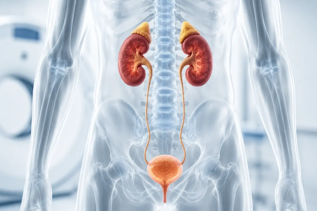
CT of the kidneys and urinary system is a layered X-ray examination that helps assess the kidneys, adrenal glands, urinary tract and bladder. Computed tomography may be prescribed to detect stones, clarify the causes of blood in the urine, evaluate cysts, inflammatory changes, neoplasms, impaired urine outflow and other conditions.
At the HBMedical medical center, you can undergo a kidney CT in Kyiv without contrast or with intravenous contrast enhancement. The examination is performed on a Siemens SOMATOM go.Up CT scanner — 32 slices and up to 64 slices in IVR mode, with the option of using low-dose protocols. The center is located at: 3 Oleksandra Dovzhenka Street, Kyiv, near Shuliavska metro station.
In the HBMedical price list, the examination is presented as a CT of the urological complex, which includes the kidneys, adrenal glands and bladder. A separate non-contrast protocol covering the kidneys, urinary tract and bladder is available for stone detection.
You can book a CT of the urinary system by phone or through the form on the website. When booking, provide the exact wording from the referral and specify which examination is required: the urological complex, contrast-enhanced kidney CT, bladder CT or stone detection.
Price of kidney and urinary system CT in Kyiv
According to the current HBMedical price list, the examination costs:
- CT of the urological complex — kidneys, adrenal glands and bladder without contrast: UAH 2,500;
- CT of the kidneys, adrenal glands and bladder with contrast: UAH 4,900;
- CT for detecting stones in the kidneys, urinary tract and bladder without contrast: UAH 2,500.
The examination price includes a radiologist’s report and a USB flash drive with the results. The price list has been valid since April 1, 2026, but it is recommended to confirm the current price before your visit.
The required protocol depends on the clinical task. CT without contrast is usually used to detect stones. To assess kidney masses, inflammatory changes, the urinary tract and bladder, the doctor may prescribe a contrast-enhanced examination.
What is a CT of the urinary system?
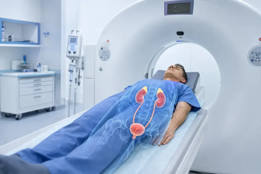
The urinary system is responsible for producing, storing and excreting urine. It includes:
- the kidneys;
- the renal calyces and pelvises;
- the ureters;
- the bladder;
- the urethra.
The adrenal glands are not directly part of the urinary system, but they are located above the kidneys and are often evaluated during the same CT examination.
Computed tomography uses X-rays and computer processing to create a series of thin cross-sectional images. They can be viewed in different planes and used to generate three-dimensional reconstructions. CT helps assess organs, blood vessels, surrounding fatty tissue and bone structures at the same time.
What is included in the urological CT complex?
According to the HBMedical price list, the urological complex covers:
- the right and left kidneys;
- the pelvicalyceal system;
- the right and left adrenal glands;
- the bladder;
- the perirenal fat;
- retroperitoneal lymph nodes within the scanning area;
- adjacent blood vessels and other anatomical structures.
The ureters may be assessed within the scanning area, but the quality of their visualization depends on the protocol, filling of the urinary tract and contrast phases.
If the referral specifies CT of the ureters, CT of the urinary tract or CT urography, be sure to mention this when booking. Not every contrast-enhanced kidney CT is CT urography: urography requires a special protocol with a phase during which the contrast is excreted by the kidneys and fills the urinary tract. CT urography is used to assess the kidneys, ureters and bladder, including in patients with hematuria or suspected urinary tract disease.
What does a kidney CT show?
Computed tomography helps assess the location, size, shape and structure of the kidneys.
A kidney CT may show:
- stones;
- dilatation of the pelvicalyceal system;
- hydronephrosis;
- impaired urine outflow;
- cysts;
- complex cystic lesions;
- benign and suspicious tumors;
- angiomyolipoma;
- inflammatory changes;
- a kidney abscess;
- changes in the perirenal fat;
- traumatic injury;
- hemorrhage;
- congenital features;
- abnormal kidney position or fusion;
- postoperative changes;
- the condition after tumor treatment;
- possible spread of a pathological process to adjacent structures.
For detailed characterization of an indeterminate kidney lesion, CT before and after intravenous contrast administration is often used. Comparing the phases helps assess contrast enhancement and the blood supply characteristics of the lesion.
CT does not always allow the exact tumor type to be determined. MRI, laboratory tests, follow-up imaging or biopsy may be required to establish the diagnosis.
Kidney CT with contrast
A contrast-enhanced kidney CT is performed with intravenous administration of an iodine-containing agent. The contrast reaches the kidneys through the bloodstream, after which it is filtered and excreted through the urinary tract.
A contrast-enhanced examination helps:
- assess the blood supply to the kidneys;
- distinguish a cyst from a solid lesion;
- clarify the structure of a detected tumor;
- assess the margins of a neoplasm;
- determine vascular involvement;
- evaluate the renal pelvises and ureters;
- detect an inflammatory process;
- assess an abscess;
- investigate the consequences of trauma;
- determine the spread of a pathological process;
- perform follow-up after surgery or other treatment.
To assess kidney lesions, the doctor may prescribe multiphase scanning: before contrast administration and during the vascular, nephrographic or excretory phase. The exact set of phases depends on the clinical task.
Kidney CT without contrast
Non-contrast CT is particularly useful for detecting stones in the kidneys and urinary tract. The method helps determine:
- whether a stone is present;
- its size;
- its exact location;
- the number of calculi;
- the stone density;
- the level of ureteral obstruction;
- dilatation of the urinary tract above the obstruction;
- associated changes around the kidney.
HBMedical offers a separate examination for this purpose — CT for detecting calculi in the kidneys, urinary tract and bladder. It is performed without intravenous contrast enhancement and costs UAH 2,500.
Non-contrast CT may also provide some information about kidney size, cysts, hemorrhage and certain lesions. However, contrast enhancement may be required for a complete characterization of many pathological changes.
CT for kidney stones
CT may be prescribed when urolithiasis is suspected, especially if the patient has:
- sharp pain in the side or lower back;
- pain radiating to the groin;
- nausea or vomiting;
- blood in the urine;
- painful urination;
- frequent urges to urinate;
- suspected ureteral obstruction;
- recurrent attacks of renal colic.
Non-contrast CT is often used for the initial detection of stones. Contrast may make some small calculi more difficult to detect, so the protocol should be selected according to the referral.
If severe pain is accompanied by a high fever, chills, marked weakness or an inability to urinate, urgent medical consultation is required.
CT of the adrenal glands
The adrenal glands are small glands located above the upper poles of the kidneys. They produce hormones involved in regulating blood pressure, metabolism and the body’s response to stress.
CT of the adrenal glands may be prescribed for:
- an incidentally detected lesion;
- suspected adenoma;
- hormonal disorders;
- high blood pressure of unclear origin;
- suspected pheochromocytoma;
- suspected malignant lesion;
- the need to assess metastatic involvement;
- follow-up of a previously detected nodule;
- preoperative planning.
CT helps assess the size, density, margins and structure of a lesion. To clarify the nature of a nodule, a special protocol before and after contrast administration may be used to assess contrast uptake and washout. At the same time, a non-contrast examination is sufficient for some lesions, so the protocol is determined by the doctor.
CT shows anatomical changes but does not measure hormone levels. If an adrenal lesion is present, laboratory tests and an endocrinologist’s consultation may also be required.
What does a bladder CT show?
During a bladder CT, the doctor can assess:
- the shape and size of the organ;
- wall thickness;
- uniformity of filling;
- stones;
- diverticula;
- space-occupying lesions;
- suspicious wall thickening;
- inflammatory changes in the surrounding tissues;
- blood clots;
- the condition of adjacent organs;
- enlarged lymph nodes;
- the spread of an established tumor process;
- postoperative changes.
Contrast-enhanced bladder CT may help identify a suspicious area and assess the spread of the process. CT urography is also used in cases of blood in the urine to assess the kidneys, ureters and bladder.
Computed tomography does not always detect very small superficial changes of the mucous membrane. If a bladder tumor is suspected, the urologist may additionally prescribe cystoscopy, urine tests or biopsy.
Bladder CT with contrast
Contrast enhancement helps:
- assess the bladder wall;
- visualize a suspicious lesion more clearly;
- determine its size;
- evaluate the perivesical fat;
- check the pelvic lymph nodes;
- assess contrast excretion by the kidneys;
- visualize the ureters during the excretory phase;
- determine the extent of an already diagnosed disease.
For a high-quality examination, the bladder may need to be moderately full. The patient may be asked to drink water and avoid urinating until the scan is completed. The exact instructions depend on the medical center’s protocol.
Bladder CT in women and men
The CT procedure is performed in the same way for women and men. However, different adjacent organs may be included in the scanning area.
In men, the examination may show:
- the prostate gland;
- the seminal vesicles;
- the surrounding pelvic tissues.
In women, it may partially assess:
- the uterus;
- the adnexa;
- the surrounding pelvic tissues.
If a targeted examination of the pelvic organs, prostate gland or female reproductive organs is required, a separate CT or MRI protocol may be needed.
Renal angiomyolipoma on CT
Angiomyolipoma is a kidney lesion that may contain fatty tissue, blood vessels and muscle components. The presence of visible fat often helps suggest this type of lesion on CT.
The radiologist evaluates:
- the size of the angiomyolipoma;
- its location;
- the number of lesions;
- its relationship with the pelvicalyceal system and blood vessels;
- the presence of signs of hemorrhage;
- changes over time.
Not all fat-containing lesions have the same nature, and some angiomyolipomas may contain little visible fat. Therefore, the CT report should be shown to a urologist.
Oncocytoma and kidney cancer on CT
Contrast-enhanced CT helps assess a suspicious kidney lesion, including its size, margins, structure, blood supply and relationship with blood vessels.
CT can be used to assess:
- the location of the tumor;
- extension beyond the kidney;
- involvement of the renal vein;
- the condition of the perirenal fat;
- regional lymph nodes;
- adjacent organs;
- postoperative changes.
Oncocytoma is a benign lesion, but on CT it may sometimes resemble renal cell carcinoma. Therefore, final determination of the tumor type may require histological examination. CT helps characterize the lesion and plan further diagnostics, but it does not always replace biopsy or surgical pathology.
When is a CT of the urinary system prescribed?
A referral for the examination may be issued by a urologist, nephrologist, oncologist, endocrinologist, surgeon or family doctor.
Possible indications include:
- lower back or side pain;
- renal colic;
- blood in the urine;
- recurrent urinary tract infections;
- impaired urine outflow;
- suspected stone;
- hydronephrosis;
- suspected kidney cyst or tumor;
- changes detected on ultrasound;
- suspected bladder neoplasm;
- suspicious thickening of the bladder wall;
- trauma to the kidneys or urinary system;
- follow-up after surgery;
- follow-up after tumor treatment;
- congenital abnormalities;
- an adrenal lesion;
- the need to clarify laboratory, ultrasound or MRI results.
Blood in the urine may have different causes — from an infection or stone to a more serious disease, so it should not be ignored. To assess hematuria, the doctor may prescribe CT urography, ultrasound, cystoscopy and laboratory tests depending on the clinical situation.
CT urography and standard kidney CT: what is the difference?
Standard kidney CT
It may be performed without contrast or during a single contrast phase. The protocol depends on whether the goal is to detect a stone, assess inflammation or examine a kidney lesion.
CT urography
This is a specialized examination of the kidneys, ureters and bladder after intravenous contrast administration. It includes an excretory phase during which the contrast enters the urine and fills the urinary tract.
CT urography may be used for:
- visible blood in the urine;
- suspected ureteral tumor;
- the need to assess the pelvicalyceal system;
- recurrent infections with a complicated course;
- postoperative changes;
- congenital abnormalities of the urinary tract.
If the referral specifically states CT urography, inform the administrator before booking so that the correct protocol can be arranged.
Preparation for kidney CT without contrast
Complex preparation is usually not required for non-contrast stone detection.
Before the examination, it is recommended to:
- bring the doctor’s referral;
- bring previous CT, MRI and ultrasound examinations;
- bring laboratory test results and medical records;
- report a possible pregnancy;
- report any recent examinations involving radiation;
- clarify whether the bladder needs to be filled;
- remove metal objects from the scanning area;
- not stop prescribed medications on your own.
Exact instructions regarding fluid intake and bladder filling should be clarified when booking.
Preparation for kidney CT with contrast
Before a contrast-enhanced kidney CT, inform the staff about:
- kidney disease;
- reduced kidney function;
- previous reactions to iodine-containing contrast agents;
- thyroid disease;
- diabetes mellitus;
- bronchial asthma;
- severe allergic reactions;
- the medications you take;
- pregnancy or the possibility of pregnancy.
To assess kidney function, a recent blood creatinine test with estimated glomerular filtration rate may be required. The validity period of the test and the result requirements should be confirmed with HBMedical.
Before a contrast-enhanced CT, the patient may be asked not to eat for a certain period. To assess the urinary tract and bladder, the patient may also be advised to drink water and temporarily avoid urinating. Preparation rules vary depending on the protocol and medical facility.
Do not stop taking medications on your own. Decisions regarding medications should be made by a doctor or medical staff.
How is a CT of the kidneys and bladder performed?
Before the examination, the patient leaves metal objects outside the scanning room. For a contrast-enhanced protocol, a healthcare professional inserts an intravenous catheter.
The procedure takes place in several stages:
- Positioning. The patient lies on the movable table of the CT scanner.
- Initial scanning. If necessary, a non-contrast series is performed first.
- Contrast administration. An iodine-containing agent is administered intravenously using an automatic injector.
- Contrast phases. The scanner performs imaging at the required times after the agent is administered.
- Excretory phase. If the ureters and bladder need to be assessed, an additional series may be performed after the kidneys excrete the contrast.
- Result processing. The radiologist analyzes the images in different planes and prepares a report.
During contrast administration, a brief feeling of warmth, a metallic taste or a sensation similar to the urge to urinate may occur. To obtain clear images, it is important to remain still and follow the staff’s instructions.
Contraindications and limitations
CT uses ionizing radiation, so the examination is performed for medical indications.
Before the procedure, report:
- pregnancy or the possibility of pregnancy;
- recent CT examinations;
- an inability to remain still;
- severe pain;
- a condition that makes it difficult to remain in a horizontal position.
For contrast-enhanced CT, the following are also considered:
- kidney function;
- previous reactions to iodine-containing agents;
- certain thyroid disorders;
- the use of certain medications;
- the patient’s general condition.
A pacemaker, orthopedic implants and most metal structures are not contraindications to CT.
CT, MRI or ultrasound of the bladder and kidneys
Ultrasound
It is often used as an initial assessment method for:
- kidney size;
- dilatation of the pelvicalyceal system;
- large cysts;
- the bladder;
- residual urine;
- certain stones.
Ultrasound does not use ionizing radiation, but it may not show a small stone in the ureter or some deeply located structures.
CT
CT is well suited for:
- detecting stones;
- assessing the ureters;
- diagnosing renal colic;
- characterizing lesions after contrast enhancement;
- assessing trauma;
- determining the spread of a tumor process;
- a comprehensive examination of the kidneys and bladder.
MRI
MRI may be used for detailed assessment of certain kidney and bladder lesions and the pelvic organs. The method does not use ionizing radiation, but it is less suitable for detecting most stones and takes longer.
The choice between CT, MRI and ultrasound depends on the symptoms, previous results and the clinical task.
What does the patient receive after CT?
After the scan is completed, the radiologist analyzes the images and prepares a report.
The patient receives:
- CT images of the kidneys and urinary system organs;
- a radiologist’s report;
- the examination recorded on a USB flash drive;
- materials for consultation with a urologist, nephrologist, oncologist, endocrinologist or another specialist.
At HBMedical, CT results are usually prepared within 5–7 hours. In complex cases, the time may vary. The report describes the detected changes, but the final diagnosis is established by the relevant specialist.
Why undergo a kidney CT at HBMedical?
- Siemens SOMATOM go.Up CT scanner;
- 32 slices and up to 64 slices in IVR mode;
- the option of using low-dose protocols;
- CT of the urological complex with or without contrast;
- a separate non-contrast protocol for stone detection;
- assessment of the kidneys, adrenal glands and bladder;
- image interpretation by a radiologist;
- the report and USB flash drive are included in the price;
- results are usually prepared within 5–7 hours;
- location near Shuliavska metro station;
- advance booking by phone or online.
Where to undergo a kidney CT in Kyiv
You can undergo CT of the kidneys, adrenal glands and urinary system at the HBMedical medical center at: 3 Oleksandra Dovzhenka Street, Kyiv, near Shuliavska metro station.
The medical center is located on the right bank of Kyiv. It is convenient to reach for patients from Shuliavka, KPI, Lukianivka, Dorohozhychi and Nyvky, as well as the Shevchenkivskyi, Solomianskyi and Sviatoshynskyi districts.
When booking, inform the administrator about:
- the exact examination name from the referral;
- whether stones need to be detected;
- whether contrast enhancement has been prescribed;
- whether CT urography is required;
- whether the bladder or adrenal glands need to be assessed specifically;
- whether previous CT, MRI or ultrasound examinations are available;
- whether you have kidney disease;
- whether you have had reactions to contrast;
- whether you have thyroid disease;
- whether you are pregnant or may be pregnant.
Book a CT of the kidneys and urinary system at HBMedical to undergo the examination in Kyiv near Shuliavska and receive the results for further consultation with your doctor.
Frequently asked questions
What does a kidney CT show?
CT helps detect stones, cysts, lesions, hydronephrosis, inflammatory changes, an abscess, traumatic injury and impaired urine outflow.
How much does a kidney CT cost in Kyiv?
According to the current HBMedical price list, the urological complex without contrast costs UAH 2,500, and with contrast enhancement — UAH 4,900.
What is included in the urological CT complex?
According to the HBMedical price list, the examination covers the kidneys, adrenal glands and bladder. The extent of ureter assessment depends on the selected protocol and contrast phases.
How much does a CT for stone detection cost?
Non-contrast CT of the kidneys, urinary tract and bladder for detecting calculi costs UAH 2,500.
When is a contrast-enhanced kidney CT needed?
Contrast may be required to assess a lesion, inflammatory process, kidney blood supply, trauma, the urinary tract and the spread of pathological changes.
Are stones visible on CT without contrast?
Yes. Non-contrast CT is one of the main methods for detecting stones in the kidneys and ureters. It helps determine the size, location and number of calculi.
What does a bladder CT show?
The examination may show stones, diverticula, wall thickening, a space-occupying lesion, inflammatory changes and the spread of an already established pathological process.
Is contrast required for a bladder CT?
Not always. Contrast is more commonly used in cases of hematuria, suspected neoplasm, the need to assess the ureters or the spread of a process.
How should I prepare for a kidney CT with contrast?
You should report kidney disease, thyroid disease and previous reactions to contrast. A creatinine test may be required. Instructions regarding food, fluid intake and bladder filling should be clarified when booking.
Does the bladder need to be full?
It depends on the protocol. To assess the bladder or perform CT urography, the patient may be asked to drink water and avoid urinating until the scan is completed.
Can a kidney CT be performed without contrast?
Yes. A non-contrast protocol is used primarily to detect stones and in certain other clinical situations.
How does CT urography differ from kidney CT?
CT urography includes a special excretory phase after contrast administration and is intended to assess the kidneys, ureters and bladder. A standard kidney CT may use a different set of phases.
Is a referral required?
You can clarify whether CT can be performed without a referral when booking. A referral is recommended because it helps select the correct examination: stone detection, the urological complex, contrast-enhanced CT or CT urography.
When will the report be ready?
At HBMedical, CT results are usually prepared within 5–7 hours. In complex cases, the report may take longer.
This material is for informational purposes only and does not replace a medical consultation.










Мені все сподобалось
Всі профі
Дякую


Велика подяка лікарю Дуброву Є.О. — надзвичайно компетентний і чуйний фахівець, який детально все пояснив, розвіяв тривогу та підтримав морально. Щиро дякую анестезіологу Бєлаш О.М. за високий професіоналізм, чуйність і справжнє людське ставлення — занурення в сон та сам перебіг процедури минули максимально комфортно. Також величезне спасибі медсестрі (на жаль, не дізналася ім'я) за її турботу, доброту та увагу.
Весь персонал вразив своєю чуйністю та теплом. Дякую за ваш труд і турботу про пацієнтів! Однозначно рекомендую.














Дуже ввічливий та приємний персонал клініки. Лікар був уважний, без поспіху пройшовся по всім аспектам здоровʼя. Лабораторія швидко зробила аналізи. В цілому дуже задоволена. Радже звертатися!




Саме до таких фахівців, в разі необхідності, є бажання повернутися!

і закінчуючи отриманим описом і рекомендаціями від лікарів.
























Need specialist help?
Order a callback and our consultant will help you choose a doctor and make an appointment at a convenient time for you!