








 Work time
Diagnostics: Mon - Sun, 6:00 - 23:00
Polyclinic: Mon - Sun, 8:00 - 20:00

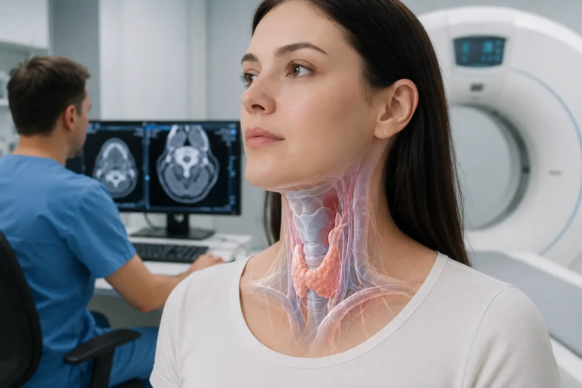
CT of the soft tissues of the neck is a cross-sectional X-ray examination of the larynx, pharynx, lymph nodes, salivary glands, thyroid gland and other anatomical structures of the neck. The examination helps detect inflammatory changes, abscesses, enlarged lymph nodes, masses and other pathological processes.
At HBMedical, patients can undergo a neck CT in Kyiv without contrast or with intravenous contrast enhancement. The examination is performed on a Siemens SOMATOM go.Up CT scanner — 32 slices and up to 64 slices in IVR mode. The centre is located at 3 Oleksandra Dovzhenka Street, Kyiv, near Shuliavska metro station.
For a detailed assessment of soft tissues, lymph nodes, inflammatory processes and masses, a doctor often orders a contrast-enhanced neck CT. The contrast agent helps distinguish blood vessels, organs and pathologically altered tissues more clearly.
You can book a CT scan of the soft tissues of the neck at HBMedical by phone or through the website form. When booking, provide the exact wording from the referral: neck CT, laryngeal CT, throat CT or contrast-enhanced CT of the soft tissues of the neck.
Price of a neck CT in Kyiv
According to the current HBMedical price list, the cost of the examination is:
- CT of the soft tissues of the neck without contrast — UAH 2,800;
- CT of the soft tissues of the neck with contrast — UAH 4,100.
In the price list, the examination is listed as «CT of the soft tissues of the neck — larynx, throat». The price includes a radiologist’s report and a copy of the examination on a USB flash drive. Before your visit, it is advisable to confirm the current price and whether contrast enhancement is required.
The choice of protocol depends on the clinical question. Contrast-enhanced neck CT is more often required when assessing lymph nodes, deep neck spaces, an inflammatory process or a suspicious mass. For certain tasks, the doctor may prescribe a non-contrast examination.
What is CT of the soft tissues of the neck?
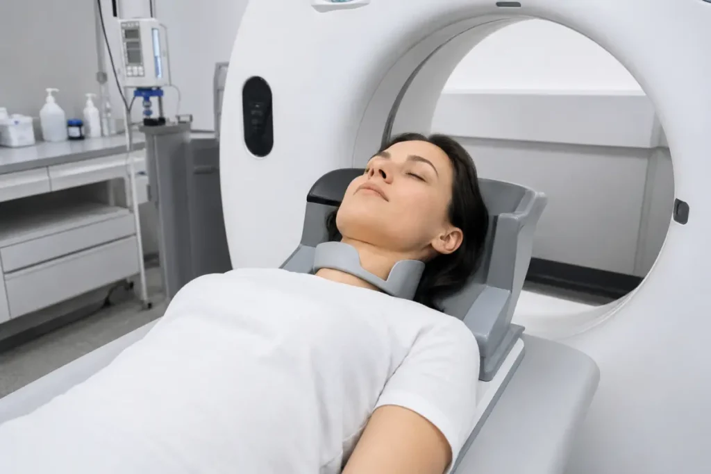
Computed tomography creates a series of cross-sectional images of the area being examined. The resulting slices can be viewed in different planes and, when necessary, used to create three-dimensional reconstructions. The method makes it possible to assess internal organs, bones, soft tissues and blood vessels.
A neck CT may cover the area from the skull base to the upper chest. The exact scan boundaries depend on the referral, symptoms and the protocol selected by the radiologist.
In everyday language, patients may refer to this examination as:
- throat CT;
- laryngeal CT;
- CT of the soft tissues of the neck;
- CT of the neck lymph nodes;
- contrast-enhanced neck CT;
- computed tomography of the neck.
The term «throat CT» is not a precise medical description. Depending on the symptoms, the doctor may need an examination of the pharynx, larynx, nasopharynx, lymph nodes or the entire neck region.
Which structures are included in a neck CT?
Within the selected protocol, the radiologist may assess:
- the nasopharynx;
- the oropharynx;
- the hypopharynx;
- the larynx;
- the epiglottis;
- the vocal and vestibular folds;
- the subglottic region;
- the upper part of the trachea;
- the soft tissues of the floor of the mouth;
- the base of the tongue;
- the palatine tonsils;
- the parotid salivary glands;
- the submandibular salivary glands;
- the thyroid gland;
- the cervical lymph nodes;
- the deep fascial spaces of the neck;
- the neck muscles;
- the subcutaneous fat;
- the major vessels within the standard protocol;
- adjacent bony structures;
- the lung apices within the scan range.
The scope of the examination may vary. A standard CT of the soft tissues of the neck is not a complete substitute for a CT of the cervical spine, CT angiography, brain MRI or a specialised chest examination.
What does a neck CT show?
CT of the soft tissues of the neck helps assess the size, shape, structure and relative position of organs. The examination may show:
- enlarged or structurally altered lymph nodes;
- inflammatory infiltrates;
- abscesses and fluid collections;
- soft-tissue swelling;
- masses of the pharynx and larynx;
- cysts and other space-occupying lesions of the neck;
- changes in the salivary glands;
- salivary duct stones within the capabilities of the protocol;
- enlargement or heterogeneity of the thyroid gland;
- spread of a pathological process into surrounding tissues;
- narrowing or displacement of the airway;
- postoperative and scar-related changes;
- consequences of trauma;
- foreign bodies;
- congenital anatomical variations;
- lesions of bony and cartilaginous structures;
- changes relevant to planning surgery or radiation therapy.
When there is a neck mass, CT helps determine its location, size and relationship to surrounding structures. However, the nature of a mass cannot always be established conclusively from CT images alone. Examination, ultrasound, MRI, endoscopy, aspiration or biopsy may also be required.
Contrast-enhanced neck CT: what does it show?
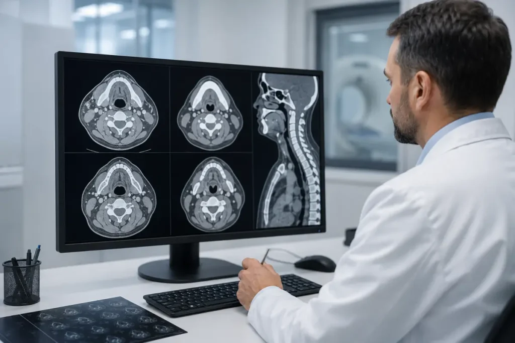
Contrast-enhanced neck CT is performed with the intravenous administration of an iodine-based agent. Contrast temporarily changes the appearance of blood vessels, organs and tissues on CT images, making pathological areas more conspicuous.
Contrast enhancement helps to:
- assess the blood supply to a mass;
- define its margins more accurately;
- distinguish blood vessels from lymph nodes;
- assess the condition of cervical lymph nodes;
- identify areas of necrosis within a mass;
- determine the spread of the process into surrounding tissues;
- assess inflammatory infiltrates;
- detect and localise an abscess;
- assess the larynx and pharynx;
- evaluate postoperative changes;
- plan further diagnosis or treatment.
For the initial assessment of a non-pulsatile neck mass, CT with intravenous contrast is among the recommended imaging methods when there are no contraindications.
Contrast should not be selected independently simply to obtain a «more accurate» result. The need for contrast is determined by the doctor, the referral and the specific clinical question.
CT of the soft tissues of the neck without contrast
A non-contrast CT may be prescribed if:
- contrast enhancement is not needed to answer the clinical question;
- calcifications or a foreign body need to be assessed;
- salivary gland stones are suspected;
- specific bony or cartilaginous structures need to be assessed;
- contrast administration is contraindicated;
- the examination is performed according to a special protocol agreed with the doctor.
Without contrast, blood vessels, lymph nodes, inflammatory changes and masses may be less clearly distinguishable from the surrounding tissues. Therefore, when a tumour, abscess or extensive inflammatory process is suspected, the doctor often prefers a contrast-enhanced examination.
When is a neck CT ordered?
A referral for CT of the soft tissues of the neck may be issued by an ENT specialist, oncologist, surgeon, endocrinologist, dentist, maxillofacial surgeon, family doctor or another specialist.
Possible indications include:
- a lump or mass in the neck;
- enlarged lymph nodes;
- neck asymmetry;
- pain and swelling;
- difficulty swallowing;
- pain when swallowing;
- a foreign-body sensation in the throat;
- persistent hoarseness;
- breathing difficulties;
- suspected inflammation of the deep neck tissues;
- suspected abscess;
- changes in the larynx or pharynx;
- suspected mass;
- a diagnosed head and neck cancer;
- the need to assess lymph nodes;
- changes in the salivary glands;
- neck trauma;
- suspected foreign body;
- preparation for surgery;
- follow-up after surgical, radiation or drug treatment;
- the need to clarify ultrasound, endoscopy, MRI or other examination findings.
Not every enlarged lymph node or throat discomfort requires CT. The choice of the initial examination depends on the patient’s age, the location of the changes, the duration of symptoms and the physical examination findings.
CT of the larynx and throat
A laryngeal CT helps assess:
- the epiglottis;
- the laryngeal vestibule;
- the vocal folds;
- the subglottic region;
- the laryngeal cartilages;
- the airway lumen;
- the surrounding soft tissues;
- the cervical lymph nodes;
- possible spread of a pathological process.
The examination may be prescribed for hoarseness, swallowing disorders, a suspected mass, consequences of trauma or the need to assess the extent of an already diagnosed condition.
CT does not replace an ENT examination of the larynx. Small superficial mucosal changes are better assessed during laryngoscopy, while computed tomography helps evaluate deep tissues, cartilage, lymph nodes and the spread of the process.
CT of the cervical lymph nodes
Lymph nodes may enlarge because of infection, inflammation and other diseases. CT helps assess:
- the location of the lymph nodes;
- their size;
- their number;
- symmetry;
- internal structure;
- the definition of their margins;
- the presence of necrotic areas;
- the condition of surrounding tissues;
- their relationship to blood vessels and organs.
Contrast enhancement helps distinguish lymph nodes from adjacent blood vessels and soft tissues more clearly. However, CT does not always allow the cause of an enlarged node to be determined conclusively. Depending on the indications, the doctor may recommend ultrasound, aspiration or biopsy.
Neck CT for inflammation and abscess
An infectious process may spread between the deep fascial spaces of the neck. Contrast-enhanced CT helps assess:
- the location of the inflammatory process;
- spread into adjacent tissues;
- the presence of a fluid collection;
- abscess formation;
- swelling of the pharynx or larynx;
- airway narrowing;
- involvement of the salivary glands;
- the condition of the lymph nodes;
- possible spread of the process into the upper chest.
If there is difficulty breathing, rapidly increasing neck swelling, inability to swallow saliva or a sudden deterioration in condition, urgent medical care is required rather than waiting for a scheduled examination.
CT of the salivary glands
During CT of the soft tissues of the neck, the parotid and submandibular salivary glands can be assessed.
The examination may help detect:
- gland enlargement;
- inflammatory changes;
- an abscess;
- a stone in a salivary gland or duct;
- a cyst;
- a space-occupying lesion;
- spread of a pathological process;
- enlarged adjacent lymph nodes.
Ultrasound may be the initial method for a superficial salivary gland mass. CT or MRI is used when it is necessary to assess the deep part of the gland, the surrounding tissues or the extent of the process.
Does CT show the thyroid gland?
The thyroid gland is included in the neck CT scan range and can be assessed for:
- size;
- location;
- symmetry;
- marked heterogeneity;
- large nodules;
- calcifications;
- displacement of the trachea;
- extension of a mass beyond the gland;
- enlarged lymph nodes.
However, a neck CT does not replace a targeted thyroid ultrasound. Ultrasound is better suited to assessing the structure of small nodules and helps select an area for aspiration. CT may be useful for large masses, retrosternal extension or when it is necessary to assess the relationship of the gland to the trachea, blood vessels and other structures.
If the patient is preparing for radioactive iodine treatment or has thyroid disease, the doctor must be informed before an iodine-containing contrast agent is administered.
Neck CT and cervical spine CT — what is the difference?
These are two different examinations.
CT of the soft tissues of the neck
The main focus is on:
- the pharynx;
- the larynx;
- the lymph nodes;
- the salivary glands;
- the thyroid gland;
- the muscles;
- the fascial spaces;
- masses and inflammatory changes.
CT of the cervical spine
It is intended to assess:
- the cervical vertebrae;
- fractures;
- bony canals;
- spinal joints;
- degenerative changes;
- the position of metal hardware.
If the referral says «neck CT», it is worth asking the doctor whether it refers to the soft tissues or the cervical spine. At HBMedical, these are separate services with different protocols and prices.
Neck CT and CT angiography of the neck vessels
A standard CT of the soft tissues of the neck can show the major blood vessels, especially after contrast administration, but it does not replace specialised CT angiography.
CT angiography is prescribed when a detailed assessment is needed of:
- the carotid arteries;
- the vertebral arteries;
- narrowing or occlusion of a vessel;
- an aneurysm;
- arterial wall dissection;
- a vascular malformation;
- a pulsatile neck mass;
- the consequences of vascular trauma.
If the mass is pulsatile, this must be reported when booking. Standard neck CT and CT angiography use different contrast administration and scanning protocols.
Neck CT, ultrasound or MRI
Ultrasound
Ultrasound is often used for the initial assessment of:
- the thyroid gland;
- superficial lymph nodes;
- superficial masses;
- the salivary glands;
- blood flow in the vessels.
The method does not use ionising radiation, but its capabilities are limited when examining deep spaces, structures behind bones and air-filled regions.
Computed tomography
CT rapidly creates cross-sectional images and provides good visualisation of:
- the deep spaces of the neck;
- the larynx and pharynx;
- bony and cartilaginous structures;
- lymph nodes;
- abscesses;
- the spread of an inflammatory or neoplastic process.
Magnetic resonance imaging
MRI does not use ionising radiation and may provide a more detailed assessment of certain soft-tissue structures, nerves and the spread of a pathological process. However, the procedure takes longer and has specific limitations related to implanted devices.
The examination method is selected by the doctor according to the clinical question. Ultrasound, contrast-enhanced CT or MRI may be used to assess a neck mass.
How to prepare for a neck CT
No special preparation is usually required for a neck CT without contrast.
Before the examination, it is advisable to:
- bring the doctor’s referral;
- bring previous CT, MRI and ultrasound studies and medical records;
- bring endoscopy or biopsy results, if available;
- report any previous surgery and treatment;
- remove chains, earrings and other metal jewellery;
- remove hearing aids;
- report any metal hardware in the head and neck region;
- report pregnancy or the possibility of pregnancy;
- not stop prescribed medication on your own.
Dental implants, fillings and fixed dentures usually do not prevent CT, but they may create artefacts on the images.
Preparation for a contrast-enhanced neck CT
Before contrast-enhanced CT of the soft tissues of the neck, the staff must be informed about:
- kidney disease;
- previous reactions to iodine-containing contrast;
- thyroid disease;
- bronchial asthma;
- severe allergic reactions;
- diabetes mellitus;
- medications taken regularly;
- pregnancy or the possibility of pregnancy.
To check kidney function before contrast administration, HBMedical may require a recent creatinine test result. The exact requirements regarding food, fluids and the validity period of the test should be confirmed when booking.
Do not stop taking medication on your own. Recommendations regarding medication should be provided by the doctor or the medical centre staff.
How is a neck CT performed?
Before the procedure, the patient removes metal objects from the head and neck area. If contrast enhancement is planned, a healthcare professional inserts an intravenous catheter.
The examination is carried out in several stages:
- Positioning. The patient lies on the moving table of the CT scanner.
- Head immobilisation. The head is placed in the correct position and, if necessary, secured with soft supports.
- Contrast administration. In a contrast-enhanced protocol, the agent is administered intravenously.
- Scanning. The table moves through the scanner ring, while the machine acquires a series of cross-sectional images of the neck.
- Image processing. The acquired slices are reconstructed in different planes.
- Report preparation. The radiologist analyses the images and prepares a written description.
During the administration of iodine-containing contrast, a brief sensation of warmth or a metallic taste may occur. Contrast improves the visibility of blood vessels, organs and tissues.
To obtain clear images, the patient must remain still and follow the staff’s instructions. The exact amount of time spent at the centre depends on the protocol and whether contrast enhancement is required.
Contraindications and limitations
CT uses ionising radiation, so the examination is performed for medical indications.
Before the procedure, the staff should be informed about:
- pregnancy or the possibility of pregnancy;
- recent CT examinations;
- a condition that makes it impossible to remain still;
- severe pain;
- breathing difficulties;
- inability to lie in a horizontal position.
For contrast-enhanced CT, kidney function, previous reactions to iodine-containing agents and certain thyroid disorders are also taken into account.
The presence of a pacemaker, orthopaedic implants or most other metal devices is not a contraindication to CT. However, metal within the scan area may reduce image quality.
What does the patient receive after CT?
After the scan is completed, the radiologist analyses the cross-sectional images and prepares a report.
The patient receives:
- CT images of the soft tissues of the neck;
- a radiologist’s report;
- a copy of the examination on a USB flash drive;
- materials for consultation with an ENT specialist, surgeon, oncologist, endocrinologist or another specialist.
At HBMedical, CT results are usually prepared within 5–7 hours. In complex cases, the time may vary. The radiologist’s report describes the detected changes but does not replace the final clinical diagnosis.
Why undergo a neck CT at HBMedical?
- Siemens SOMATOM go.Up CT scanner;
- 32 slices and up to 64 slices in IVR mode;
- the option of using low-dose protocols;
- CT of the soft tissues of the neck with and without contrast;
- assessment of the larynx, pharynx, salivary glands and lymph nodes;
- image interpretation by a radiologist;
- the report and USB flash drive are included in the price;
- results are usually prepared within 5–7 hours;
- the diagnostic department is open daily;
- location near Shuliavska metro station;
- advance booking by phone or online.
Where to have a neck CT in Kyiv
You can undergo CT of the soft tissues of the neck at HBMedical, located at 3 Oleksandra Dovzhenka Street, Kyiv, near Shuliavska metro station.
The medical centre is located on the right bank of Kyiv. It is convenient for patients from Shuliavka, KPI, Lukianivka, Dorohozhychi and Nyvky, as well as the Shevchenkivskyi, Solomianskyi and Sviatoshynskyi districts.
When booking, inform the administrator about:
- the exact name of the examination from the referral;
- the main symptoms or clinical question;
- whether contrast enhancement is required;
- whether previous CT, MRI or ultrasound studies are available;
- whether surgery or treatment has been performed;
- whether there is kidney disease;
- whether there have been reactions to iodine-containing contrast;
- whether there is thyroid disease;
- whether the patient is pregnant or may be pregnant.
Book a CT scan of the soft tissues of the neck at HBMedical to undergo the examination in Kyiv near Shuliavska metro station and receive the results for further consultation with the appropriate specialist.
Frequently asked questions
What does a neck CT show?
CT helps assess the larynx, pharynx, lymph nodes, salivary glands, thyroid gland, deep spaces and other soft tissues of the neck. The examination may show inflammation, an abscess, enlarged lymph nodes, cysts, masses and postoperative changes.
How much does a neck CT cost in Kyiv?
According to the current HBMedical price list, CT of the soft tissues of the neck without contrast costs UAH 2,800. Contrast-enhanced neck CT costs UAH 4,100.
What is included in CT of the soft tissues of the neck?
Within the protocol, the pharynx, larynx, cervical lymph nodes, salivary glands, thyroid gland, muscles, fascial spaces and adjacent anatomical structures are assessed.
Is contrast required for a neck CT?
Not always, but when a tumour, abscess, extensive inflammatory process or lymph node involvement is suspected, contrast often provides more diagnostic information. The need for contrast enhancement is determined by the doctor.
How does a contrast-enhanced neck CT differ from a standard CT?
After the intravenous administration of an iodine-containing agent, blood vessels, lymph nodes, organs and pathologically altered tissues are visualised more clearly. This helps assess the margins and extent of the process more accurately.
Does a neck CT show the larynx and throat?
Yes. The examination can assess the larynx, different parts of the pharynx and the surrounding tissues. However, CT does not replace visual inspection of the mucosa during laryngoscopy.
Are lymph nodes visible on CT?
Yes. The doctor can assess the location, size, number and structure of the cervical lymph nodes. Contrast helps distinguish them more clearly from blood vessels and other tissues.
Is the cervical spine included in a neck CT?
The vertebrae may be included in the scan range, but CT of the soft tissues of the neck does not replace a specialised CT of the cervical spine.
Is a creatinine test required?
Before contrast-enhanced CT, a test may be required to assess kidney function. The exact requirements and the validity period of the result should be confirmed when booking.
How should I prepare for a contrast-enhanced neck CT?
You should report kidney and thyroid disease, previous reactions to contrast and the medications you take. The administrator will provide instructions about eating and whether tests are required.
How is a neck CT performed?
The patient lies on the CT scanner table while the machine creates a series of cross-sectional images of the neck. In a contrast-enhanced protocol, the agent is administered intravenously. It is important to remain still during scanning.
When will the report be ready?
At HBMedical, CT results are usually prepared within 5–7 hours. In complex cases, preparation of the report may take longer.
This material is for informational purposes and does not replace a medical consultation.










Мені все сподобалось
Всі профі
Дякую


Велика подяка лікарю Дуброву Є.О. — надзвичайно компетентний і чуйний фахівець, який детально все пояснив, розвіяв тривогу та підтримав морально. Щиро дякую анестезіологу Бєлаш О.М. за високий професіоналізм, чуйність і справжнє людське ставлення — занурення в сон та сам перебіг процедури минули максимально комфортно. Також величезне спасибі медсестрі (на жаль, не дізналася ім'я) за її турботу, доброту та увагу.
Весь персонал вразив своєю чуйністю та теплом. Дякую за ваш труд і турботу про пацієнтів! Однозначно рекомендую.














Дуже ввічливий та приємний персонал клініки. Лікар був уважний, без поспіху пройшовся по всім аспектам здоровʼя. Лабораторія швидко зробила аналізи. В цілому дуже задоволена. Радже звертатися!




Саме до таких фахівців, в разі необхідності, є бажання повернутися!

і закінчуючи отриманим описом і рекомендаціями від лікарів.
























Need specialist help?
Order a callback and our consultant will help you choose a doctor and make an appointment at a convenient time for you!