








 Work time
Diagnostics: Mon - Sun, 6:00 - 23:00
Polyclinic: Mon - Sun, 8:00 - 20:00

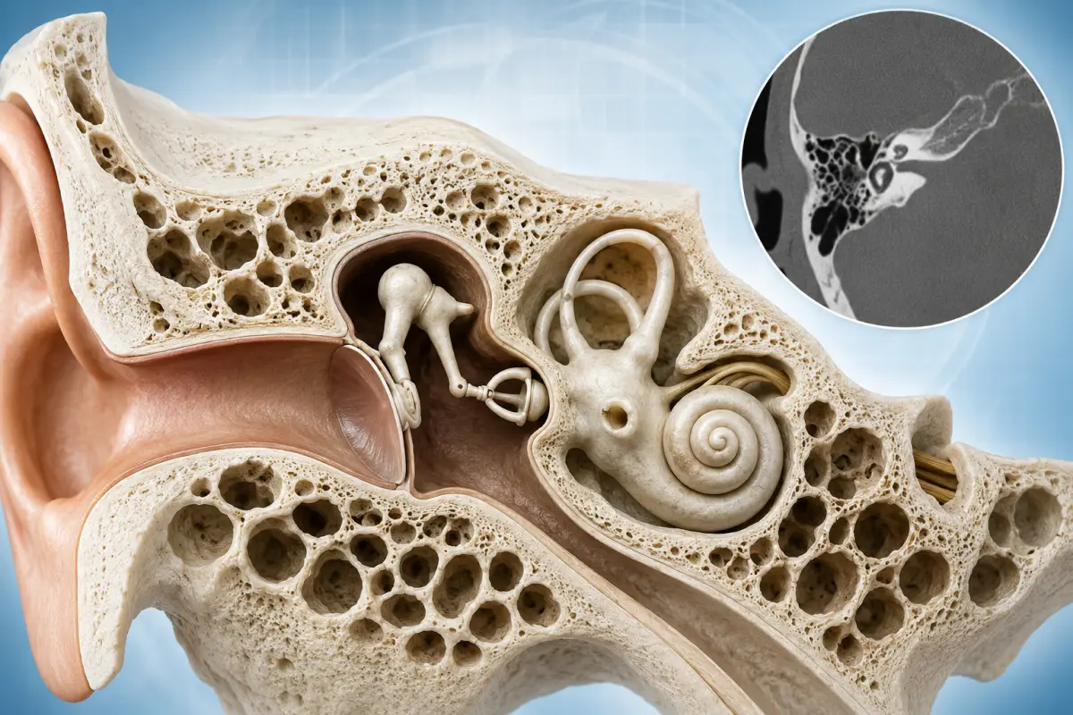
A temporal bone CT is a targeted cross-sectional examination of the structures of the outer, middle and inner ear. Computed tomography helps assess the auditory canal, tympanic cavity, auditory ossicles, mastoid process and the bony labyrinth of the inner ear in detail.
The examination may be prescribed for hearing loss, chronic otitis, suspected cholesteatoma, trauma, congenital abnormalities or before ear surgery.
At the HBMedical medical center, you can undergo a CT scan of the inner ear and temporal bones in Kyiv using a Siemens SOMATOM go.Up CT scanner with 32 slices and up to 64 slices with IVR technology.
Center address: 3 Oleksandra Dovzhenka Street, Kyiv, near the «Shuliavska» metro station.
The diagnostic department is open daily from 6:00 to 23:00. You can book an examination by phone or through the online form.
Temporal Bone CT Price in Kyiv
According to the current HBMedical price list:
- CT of the inner ear and temporal bones without contrast — UAH 2,100.
For most ear disorders, assessment of the auditory ossicles and mastoid process, fractures and congenital abnormalities, a non-contrast CT scan is used.
If the doctor has prescribed an examination with intravenous contrast, the possibility of performing it, the protocol details and the exact price should be clarified when booking.
The official price list currently contains a separate value in the «with contrast» column, but it is lower than the price of the non-contrast examination. Therefore, before publishing the price of the contrast-enhanced protocol on the page, it should be additionally confirmed with the HBMedical administration.
What Is a Temporal Bone CT?
The temporal bones are located on both sides of the skull. They contain important structures responsible for hearing and balance.
Because this area is small and anatomically complex, a special targeted CT protocol with thin slices is used. A standard brain CT does not provide the same level of detail for the middle and inner ear.
A temporal bone CT mainly allows the following bony structures to be assessed:
- the external auditory canal;
- the tympanic cavity;
- the auditory ossicles;
- the mastoid process;
- the mastoid air cells;
- the bony walls of the middle ear;
- the bony labyrinth of the inner ear;
- the cochlea;
- the vestibule;
- the semicircular canals;
- the internal auditory canal;
- the facial nerve canal;
- the carotid canal;
- the jugular fossa;
- the roof of the tympanic cavity;
- the petrous portion of the temporal bone.
A targeted temporal bone CT shows the small bony details required for diagnosing ear disorders better than a standard head CT.
CT of the Middle and Inner Ear
The terms «inner ear CT», «middle ear CT» and «temporal bone CT» are often used as closely related search terms. However, during one targeted examination, the doctor evaluates a set of structures of the outer, middle and inner ear.
Middle ear
The structures of the middle ear include:
- the tympanic cavity;
- the malleus;
- the incus;
- the stapes;
- the auditory tube within the limits of available visualization;
- the entrance to the mastoid antrum;
- the bony walls of the tympanic cavity.
CT is particularly informative for assessing the ossicular chain, chronic otitis media, cholesteatoma, destruction of the bony walls and postoperative changes.
Inner ear
The inner ear contains structures responsible for hearing and balance. CT provides the best assessment of the bony labyrinth:
- the cochlea;
- the vestibule;
- the semicircular canals;
- the vestibular aqueduct;
- the internal auditory canal.
The soft-tissue and neural structures of the inner ear are evaluated in greater detail with MRI. Therefore, the term «inner ear CT» does not mean that tomography directly shows the auditory nerve or all microscopic structures of the hearing organ.
What Does a Temporal Bone CT Show?
Computed tomography can help detect or clarify:
- acute and chronic inflammatory changes in the middle ear;
- mastoiditis;
- cholesteatoma and the bone destruction it causes;
- impaired pneumatization of the mastoid process;
- sclerotic changes in the mastoid air cells;
- damage to the auditory ossicles;
- disruption or displacement of the ossicular chain;
- otosclerosis;
- congenital abnormalities of the outer, middle or inner ear;
- narrowing or atresia of the external auditory canal;
- temporal bone fractures;
- damage to the bony facial nerve canal;
- defects in the roof of the tympanic cavity;
- semicircular canal dehiscence;
- enlargement of the vestibular aqueduct;
- bony changes around the internal auditory canal;
- foreign bodies;
- benign and suspicious bone lesions;
- postoperative changes;
- the condition after placement of a hearing implant;
- anatomical features before surgery.
CT does not measure hearing acuity and cannot independently determine the cause of every case of hearing loss. The findings should be evaluated together with an ENT examination, audiometry, tympanometry and other tests.
Middle Ear CT for Otitis
Standard acute otitis media is most often diagnosed during an ENT examination and does not always require computed tomography.
A middle ear CT may be prescribed for:
- persistent or recurrent otitis;
- chronic ear discharge;
- hearing loss;
- suspected complications;
- ineffective treatment;
- suspected mastoiditis;
- suspected cholesteatoma;
- preparation for surgery;
- the need to assess the auditory ossicles;
- suspected spread of inflammation to the bony structures.
On CT, the doctor may see filling of the tympanic cavity or mastoid air cells, impaired pneumatization, destruction of bony walls and other changes.
CT for Mastoiditis
The mastoid process is located behind the ear and contains a system of air cells connected to the middle ear. Inflammation of these structures is called mastoiditis.
CT can help assess:
- filling of the mastoid air cells;
- reduced pneumatization;
- destruction of bony septa;
- the spread of the inflammatory process;
- possible abscess formation;
- the condition of adjacent bony structures;
- the presence of intracranial or other complications within the scope of the protocol.
Acute pain behind the ear, high fever, redness, swelling or protrusion of the auricle requires urgent medical attention.
Cholesteatoma on CT
Cholesteatoma is an abnormal accumulation of epithelial tissue in the middle ear or mastoid process. It may gradually damage the surrounding bony structures.
When cholesteatoma is suspected, CT helps assess:
- the location of abnormal tissue;
- the condition of the auditory ossicles;
- destruction of the walls of the tympanic cavity;
- the roof of the middle ear;
- the facial nerve canal;
- the semicircular canals;
- the mastoid process;
- the anatomy before surgery.
However, on CT images, soft-tissue content cannot always be reliably distinguished from inflammatory fluid or granulation tissue. In complex cases, the doctor may additionally prescribe a specialized MRI.
If cholesteatoma is suspected to have spread into the inner ear or cranial cavity, both temporal bone CT and MRI of the head and internal auditory canals may be appropriate.
CT for Hearing Loss
The causes of hearing loss are conventionally divided into conductive, sensorineural and mixed.
Conductive hearing loss
This occurs when sound does not pass adequately through the outer or middle ear. Possible causes include:
- changes in the auditory ossicles;
- chronic otitis;
- cholesteatoma;
- otosclerosis;
- congenital abnormalities;
- consequences of trauma;
- postoperative changes.
When conductive hearing loss is suspected, a non-contrast temporal bone CT is one of the main imaging methods because it clearly shows the auditory ossicles and other small bony structures of the middle ear.
Sensorineural hearing loss
This may be related to the inner ear, the auditory nerve or the central auditory pathways. In such situations, the doctor more often prescribes MRI of the internal auditory canals and cerebellopontine angles.
Mixed hearing loss
This combines conductive and sensorineural components. Both CT and MRI may be needed for a complete diagnosis.
Inner Ear CT for Dizziness
Dizziness can have many causes and is not always related to a temporal bone disorder.
An inner ear CT may be prescribed if the doctor suspects:
- a congenital abnormality of the bony labyrinth;
- superior semicircular canal dehiscence;
- consequences of trauma;
- a temporal bone fracture;
- postoperative changes;
- another bony disorder of the inner ear.
For peripheral dizziness, a non-contrast temporal bone CT may be appropriate, but in many cases MRI of the head and internal auditory canals is more informative. The choice of method depends on the symptoms and the findings of neurological and ENT examinations.
Temporal Bone CT After Trauma
A temporal bone fracture may occur after a severe blow to the head, a fall, a road traffic accident or another significant injury.
CT helps assess:
- the location and direction of the fracture;
- the external auditory canal;
- the tympanic cavity;
- the ossicular chain;
- the mastoid process;
- the bony labyrinth;
- the internal auditory canal;
- the facial nerve canal;
- the carotid canal;
- the jugular fossa;
- the presence of air or fluid in abnormal areas.
Indications for a targeted CT after trauma may include:
- bleeding from the ear;
- sudden hearing loss;
- tinnitus;
- dizziness;
- balance problems;
- weakness of the facial muscles;
- leakage of clear fluid from the ear;
- suspected injury to the auditory ossicles.
In severe head trauma, the doctor determines the scope of the urgent examination. Brain CT and targeted temporal bone CT are different protocols.
CT Before Cochlear Implantation
Before cochlear implant placement, the doctor needs to assess the anatomy of the temporal bone and inner ear.
CT can help determine:
- the structure of the cochlea;
- the number and shape of its turns;
- the condition of the semicircular canals;
- the width of the internal auditory canal;
- the position of the facial nerve canal;
- congenital abnormalities;
- the consequences of inflammation or surgery;
- the condition of the mastoid process;
- features important for the surgical approach.
When planning cochlear implantation, non-contrast temporal bone CT and MRI may complement each other: CT shows the bony anatomy, while MRI shows neural and soft-tissue structures.
Contrast-Enhanced Temporal Bone CT
Contrast is generally not required to assess the bony labyrinth, auditory ossicles, chronic otitis, fractures, otosclerosis and congenital abnormalities.
Intravenous contrast may be considered in certain situations:
- when a tumor is suspected;
- in extensive inflammation;
- when complications are suspected;
- for soft-tissue assessment;
- when suspicious changes have been found during another examination.
For many middle and inner ear disorders that require assessment of soft tissues or the auditory nerve, the doctor may prescribe MRI instead of contrast-enhanced CT.
According to ACR recommendations, for conductive hearing loss and most tasks related to bony anatomy, the primary examination is temporal bone CT without intravenous contrast.
CT or MRI of the Inner Ear
CT and MRI are not interchangeable examinations.
CT is better at showing
- the auditory ossicles;
- the bony labyrinth;
- the external auditory canal;
- the mastoid process;
- fractures;
- destruction of bony structures;
- otosclerosis;
- congenital bony abnormalities;
- semicircular canal dehiscence;
- the anatomy before surgery.
MRI is better suited for assessing
- the auditory and vestibular nerves;
- the internal auditory canals;
- the cerebellopontine angles;
- soft-tissue lesions;
- inflammatory involvement of neural structures;
- sensorineural hearing loss;
- certain causes of tinnitus and dizziness;
- possible intracranial spread of a pathological process.
CT is more commonly used for conductive hearing loss, while MRI is more often used for sensorineural hearing loss. For mixed hearing loss, the doctor may prescribe both methods.
Temporal Bone CT and Brain CT — What Is the Difference?
Brain CT is primarily intended to assess:
- brain tissue;
- intracranial hemorrhage;
- the consequences of trauma;
- the ventricular system;
- large pathological lesions;
- the bones of the cranial vault within a general protocol.
Temporal bone CT is a specialized examination with thin slices and dedicated reconstructions. It allows the small structures of the middle and inner ear to be assessed in greater detail.
If the referral states «temporal bone CT», it should not be replaced independently with a standard head CT.
Indications for Temporal Bone CT
A referral for the examination may be issued by an otolaryngologist, otologist, audiologist, neurologist, neurosurgeon or another specialist.
Main indications include:
- hearing loss;
- conductive hearing loss;
- mixed hearing loss;
- chronic otitis media;
- recurrent ear inflammation;
- persistent ear discharge;
- suspected cholesteatoma;
- suspected mastoiditis;
- otosclerosis;
- noise or pulsation in the ear;
- dizziness when a bony abnormality is suspected;
- trauma to the temporal region;
- suspected fracture;
- facial muscle weakness after trauma or inflammation;
- a congenital ear abnormality;
- preparation for hearing restoration surgery;
- preparation for cochlear implantation;
- assessment of postoperative changes;
- checking implant position;
- the need to clarify audiometry, MRI or other examination results.
Preparation for Temporal Bone CT
Special preparation is usually not required for a non-contrast CT scan of the inner ear and temporal bones.
Before the examination, it is recommended to:
- bring the doctor’s referral;
- bring audiometry and tympanometry results;
- bring previous CT or MRI scans;
- bring medical records after ear surgery;
- report an implanted hearing device or cochlear implant;
- report a possible pregnancy;
- remove earrings and piercings;
- remove the hearing aid;
- remove glasses, hairpins and other metal objects;
- inform the staff if pain or another condition makes it difficult to remain still.
A hearing implant and other metal devices usually do not prevent CT, but they may create artifacts on the images.
If the doctor has prescribed contrast enhancement, a blood creatinine test and special preparation may also be required. The exact requirements should be clarified when booking.
How Is an Inner Ear CT Performed?
The examination is carried out in several stages:
- Preparation. The patient removes metal objects from the head area and completes the necessary documents.
- Positioning. The patient lies on the movable CT table. The head is placed in the correct position.
- Stabilization. Soft supports may be used if necessary to prevent movement during scanning.
- Scanning. The table moves through the scanner ring while the system creates thin cross-sectional images of the right and left temporal bones.
- Reconstruction. The acquired images are processed in different planes suitable for evaluating the middle and inner ear.
- Report preparation. The radiologist analyzes all slices and prepares a written report.
The procedure is painless. It is important not to move the head during scanning, because even slight movement can reduce the detail of the small ear structures.
Interpreting a Temporal Bone CT
After the examination, the patient receives a radiologist’s report. It may contain specialized terms.
Pneumatization of the mastoid process
This indicates how well the mastoid air cells are developed and filled with air. Pneumatization may be normal, reduced or sclerotic.
Sclerotic type of structure
This means that there are few air cells and bone tissue predominates. This may be an anatomical feature or the result of a long-term inflammatory process.
Air cells filled with soft-tissue content
This wording may indicate fluid, inflammatory material, granulation tissue or another process. The significance of the finding is determined together with the symptoms and examination findings.
The ossicular chain is intact
The malleus, incus and stapes are in the correct position without visible destruction or displacement.
Erosion of the auditory ossicles
This means damage to or destruction of part of the auditory ossicles. Such a change may occur with cholesteatoma or a chronic inflammatory process.
The bony labyrinth is unchanged
The cochlea, vestibule and semicircular canals show no visible bony abnormalities within the capabilities of CT.
Dehiscence
This means a localized absence or marked thinning of the bony wall over a particular anatomical structure, such as a semicircular canal.
The internal auditory canals are symmetrical
This means that their width and shape show no obvious asymmetry. CT alone is not sufficient to assess the auditory nerves — MRI is prescribed if necessary.
A diagnosis should not be made based on a single term in the report. The results should be shown to the ENT doctor, otologist or another specialist who referred the patient for the examination.
What Does a Normal Temporal Bone CT Look Like?
A normal CT appearance may include:
- symmetrical external auditory canals;
- air-filled tympanic cavities;
- an intact ossicular chain;
- no abnormal soft-tissue content;
- intact bony walls;
- a normally formed bony labyrinth;
- unchanged internal auditory canals;
- no fractures or areas of bone destruction;
- intact facial nerve canals;
- symmetrical or individually variable pneumatization of the mastoid processes.
Mild asymmetry or an anatomical variation does not necessarily indicate disease. The final assessment is made by the radiologist after reviewing all images.
Contraindications and Limitations
CT uses ionizing radiation, so the examination is performed when medically indicated.
Before the procedure, inform the staff about:
- pregnancy or the possibility of pregnancy;
- recent CT examinations;
- an inability to remain still;
- severe pain;
- a condition that makes head stabilization difficult.
A pacemaker, orthopedic devices and most other metal implants are not contraindications to CT.
If contrast is used, the following are also considered:
- kidney function;
- previous reactions to iodine-containing agents;
- thyroid disease;
- diabetes mellitus;
- the medications the patient is taking.
What Does the Patient Receive After CT?
After scanning, the radiologist analyzes the cross-sectional images of the temporal bones and prepares a report.
The patient receives:
- CT images of the middle and inner ear;
- a radiologist’s report;
- digital examination results;
- materials for consultation with an ENT doctor, otologist, audiologist, neurologist or another specialist.
At HBMedical, CT results are usually prepared within 5–7 hours. In complex cases, preparation of the report may take longer.
Why Choose HBMedical for a Temporal Bone CT?
- modern Siemens SOMATOM go.Up CT scanner;
- 32 slices and up to 64 slices with IVR technology;
- targeted examination of the middle and inner ear;
- assessment of the auditory ossicles and bony labyrinth;
- examination of the right and left temporal bones;
- detailed multiplanar reconstructions;
- image interpretation by a radiologist;
- the option to compare the findings with previous examinations;
- results are usually prepared within 5–7 hours;
- the diagnostic department is open daily;
- location near the «Shuliavska» metro station;
- advance booking by phone or online.
Where to Get a Temporal Bone CT in Kyiv
You can undergo a CT scan of the inner ear and temporal bones at the HBMedical medical center at 3 Oleksandra Dovzhenka Street, Kyiv, near the «Shuliavska» metro station.
The location is convenient for patients from Shuliavka, KPI, Lukianivka, Dorohozhychi, Nyvky and the Shevchenkivskyi, Solomianskyi and Sviatoshynskyi districts of Kyiv.
When booking, tell the administrator:
- the exact name of the examination stated in the referral;
- the main symptoms;
- which ear is affected;
- whether there has been an injury;
- whether any operations have been performed;
- whether a hearing implant or cochlear implant is present;
- whether previous CT scans, MRI scans or audiograms are available;
- whether contrast enhancement has been prescribed;
- whether there is a pregnancy or a possibility of pregnancy.
Schedule a temporal bone CT at HBMedical to undergo a targeted examination of the middle and inner ear in Kyiv near Shuliavska and receive the results for consultation with a doctor.
Frequently Asked Questions
What does a temporal bone CT show?
CT helps assess the external auditory canal, tympanic cavity, auditory ossicles, mastoid process and the bony labyrinth of the inner ear. The examination may show inflammatory changes, cholesteatoma, otosclerosis, fractures and congenital abnormalities.
How much does a temporal bone CT cost in Kyiv?
According to the current HBMedical price list, a non-contrast CT scan of the inner ear and temporal bones costs UAH 2,100.
Are both ears included in the examination?
During a standard targeted CT, the right and left temporal bones are usually evaluated for comparison. The exact scope should be confirmed when booking.
Is contrast required?
Contrast is generally not required to assess bony anatomy, the auditory ossicles, otosclerosis, fractures and most inflammatory changes. If a tumor or complex soft-tissue changes are suspected, the doctor may prescribe another protocol or MRI.
Does CT show the inner ear?
CT clearly shows the bony labyrinth of the inner ear: the cochlea, vestibule and semicircular canals. The auditory nerves and other soft-tissue structures are better assessed with MRI.
Can otitis be seen on CT?
CT may show abnormal content in the middle ear and mastoid air cells, but it is not always required for standard acute otitis. It is more often prescribed for a chronic process or suspected complications.
Can cholesteatoma be seen on CT?
CT helps detect soft-tissue content and characteristic destruction of bony structures. However, it does not always make it possible to distinguish cholesteatoma accurately from other inflammatory tissue.
Which is better — CT or MRI of the inner ear?
CT is better at showing bones, auditory ossicles and the bony labyrinth. MRI is better for assessing the auditory nerves, internal auditory canals and soft tissues. The method is selected according to the suspected cause of the symptoms.
Is temporal bone CT different from head CT?
Yes. Temporal bone CT uses thinner slices and a specialized protocol for detailed evaluation of the middle and inner ear.
How should I prepare for an inner ear CT?
Special preparation is usually not required for a non-contrast examination. The hearing aid, jewelry and other metal objects should be removed from the head area.
How is a temporal bone CT performed?
The patient lies on the CT table while the head is stabilized in the correct position. The scanner creates a series of thin cross-sectional images of the right and left temporal bones.
When will the report be ready?
At HBMedical, CT results are usually prepared within 5–7 hours. In complex cases, the turnaround time may be longer.
This material is for informational purposes only and does not replace a consultation with a doctor.







Дуже ввічливий та приємний персонал клініки. Лікар був уважний, без поспіху пройшовся по всім аспектам здоровʼя. Лабораторія швидко зробила аналізи. В цілому дуже задоволена. Радже звертатися!




Саме до таких фахівців, в разі необхідності, є бажання повернутися!

і закінчуючи отриманим описом і рекомендаціями від лікарів.
























Need specialist help?
Order a callback and our consultant will help you choose a doctor and make an appointment at a convenient time for you!