








 Work time
Diagnostics: Mon - Sun, 6:00 - 23:00
Polyclinic: Mon - Sun, 8:00 - 20:00

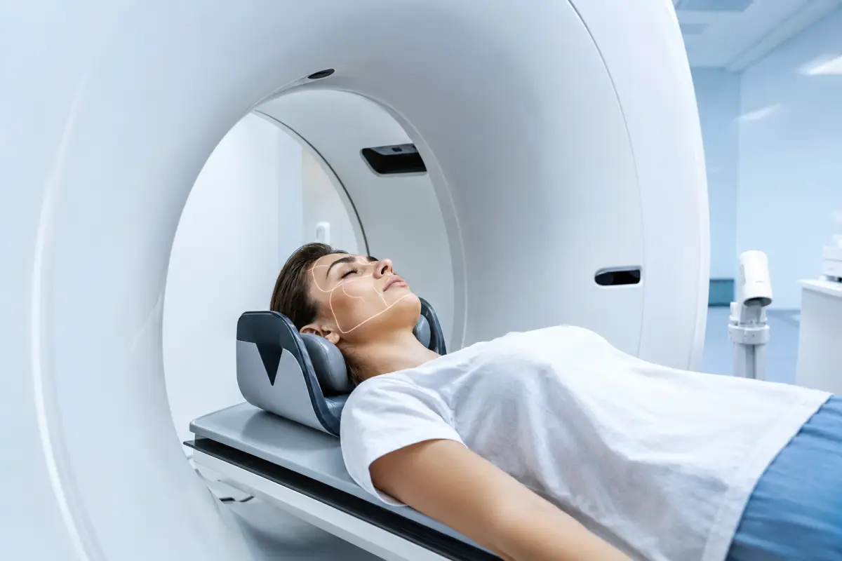
A CT scan of the facial bones is a cross-sectional X-ray examination of the bones of the face and adjacent anatomical structures. Computed tomography helps assess the complex anatomy of the facial skeleton in detail and detect fractures, displacement of bone fragments, consequences of trauma, inflammatory processes, neoplasms and other pathological changes.
At the HBMedical medical center, you can undergo a CT scan of the facial bones in Kyiv without contrast or with intravenous contrast enhancement. The examination is performed on a Siemens SOMATOM go.Up CT scanner manufactured in 2021 — 32 slices and up to 64 slices in IVR mode, with the option of using low-dose protocols.
Center address: 3 Oleksandra Dovzhenka Street, Kyiv, near Shuliavska metro station.
You can book a CT scan of the facial skeleton by phone or through the online form. When booking, state the exact wording from the referral and specify whether the paranasal sinuses, brain, orbits or skull base also need to be examined.
Price of a CT scan of the facial bones in Kyiv
HBMedical offers the following examination options:
- CT scan of the facial bones without contrast — UAH 1,700;
- CT scan of the facial bones with contrast enhancement — UAH 3,400;
- CT scan of the facial bones and paranasal sinuses with 3D reconstruction without contrast — UAH 2,400;
- CT scan of the facial bones and paranasal sinuses with 3D reconstruction and contrast enhancement — UAH 4,100.
According to the current HBMedical price list, the examination fee includes a radiologist’s report and recording of the results on a USB flash drive. The price list is valid from April 1, 2026, but patients are advised to confirm the current price before their visit.
For the assessment of fractures, deformities and most other changes in the facial bones, CT is usually performed without contrast. Intravenous contrast is mainly required when the doctor also needs to evaluate soft tissues, an inflammatory process, a neoplasm or complications.
What is a CT scan of the facial bones?
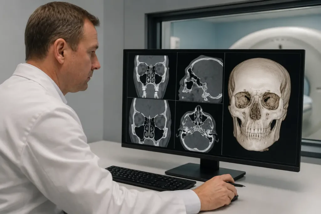
The facial skull, or facial skeleton, is a group of bones that form the contours of the face, the walls of the orbits, the nasal cavity, the upper and lower jaws, the hard palate and part of the paranasal sinuses.
Because of the complex three-dimensional anatomy of this area, a conventional X-ray does not always make it possible to determine the exact location and extent of damage. CT produces thin cross-sectional images that can be viewed in different planes and used to create a three-dimensional reconstruction. CT shows bone structures in greater detail than conventional radiography.
The terms «CT scan of the facial bones», «CT scan of the facial skeleton» and «CT scan of the facial part of the skull» are generally used to refer to similar examinations. However, the exact scan area depends on the referral and the clinical question.
Which structures are included in a CT scan of the facial bones?
During the examination, the doctor may assess:
- the nasal bones;
- the maxilla;
- the mandible within the selected scan area;
- the zygomatic bones and zygomatic arches;
- the orbital walls;
- the lacrimal bones;
- the ethmoid bone;
- the pterygoid processes;
- the hard palate;
- the bony walls of the paranasal sinuses;
- the nasal septum;
- adjacent parts of the frontal bone;
- the temporomandibular joints within the scan area;
- the junctions between the bones of the facial skeleton;
- the facial soft tissues within the diagnostic capabilities of CT.
The exact examination coverage depends on the protocol. A CT scan of the facial bones does not always include all parts of the mandible, the skull base, the temporal bones or the brain with the level of detail required for diagnosis.
What does a CT scan of the facial bones show?
Computed tomography helps assess the shape, integrity and spatial relationship of the facial bones. The examination may show:
- fractures of the nasal bones;
- fractures of the upper or lower jaw;
- fractures of the zygomatic bone and zygomatic arch;
- damage to the orbital walls;
- complex fractures of the midface;
- the displacement and number of bone fragments;
- depressed fractures;
- asymmetry of the facial skeleton;
- consequences of improperly healed fractures;
- bone defects;
- foreign bodies;
- congenital anatomical variations and deformities;
- inflammatory changes in the bones;
- signs of osteomyelitis;
- cysts and other bone lesions;
- benign and suspicious neoplasms;
- destruction or remodeling of bone tissue;
- postoperative changes;
- the condition of fixation hardware after surgical treatment;
- changes relevant to surgical planning.
CT is particularly informative for evaluating bone. For a detailed examination of the nerves, muscles, ligaments, brain or the spread of a pathological process into soft tissues, the doctor may additionally prescribe MRI.
CT scan of the facial bones after trauma
Facial trauma may result from a fall, blow, road traffic accident, sports injury or workplace accident. Because of the complex anatomy of the facial skeleton, a single impact may sometimes damage several bones.
A CT scan of the facial skeleton may be prescribed if the following symptoms occur after an injury:
- severe facial pain;
- swelling and bruising;
- deformity of the nose, jaw or cheekbone area;
- facial asymmetry;
- malocclusion;
- limited mouth opening;
- numbness of the cheek, upper lip or chin;
- double vision;
- restricted eye movement;
- sunken appearance of the eye;
- bleeding from the nose or mouth;
- clear fluid leaking from the nose;
- mobility of bone fragments;
- suspected foreign body;
- persistent symptoms despite a normal X-ray result.
For the initial assessment of suspected facial bone fractures, the ACR considers non-contrast CT of the facial bones an appropriate imaging method. Contrast is usually not required for uncomplicated trauma.
If the injury was accompanied by loss of consciousness, vomiting, seizures, severe headache or other neurological symptoms, a brain CT scan may also be required. Facial bone CT and head CT are complementary but not interchangeable examinations.
Which fractures can be detected on CT?
Computed tomography can be used to assess various types of traumatic injury:
- nasal bone fracture;
- fracture of the orbital floor;
- fracture of the medial or lateral orbital wall;
- zygomatic bone fracture;
- zygomatic arch fracture;
- maxillary fracture;
- mandibular fracture;
- Le Fort fractures;
- naso-orbito-ethmoid fracture;
- fracture of the frontal bone in the facial region;
- fractures of the walls of the paranasal sinuses;
- combined fractures involving several bones;
- old fractures and fractures that have healed incorrectly.
In the report, the radiologist may specify the location of the fracture, the direction of the fracture line, the degree of fragment displacement, involvement of the orbits or sinuses, and other features important to a traumatologist or maxillofacial surgeon.
CT scan of the facial bones with 3D reconstruction
Three-dimensional reconstruction is created from cross-sectional CT images. It helps visualize the facial bones from different angles and clearly demonstrate the spatial relationship of bone fragments, defects and deformities.
3D reconstruction may be useful for:
- complex and multiple fractures;
- displacement of bone fragments;
- orbital injuries;
- injury to the upper or lower jaw;
- deformity of the facial skeleton;
- planning maxillofacial surgery;
- reconstructive surgery;
- follow-up after the placement of plates and other fixation devices;
- the need to assess the facial bones and paranasal sinuses at the same time.
HBMedical offers a separate CT examination of the facial bones and paranasal sinuses with 3D reconstruction. The price is UAH 2,350 without contrast and UAH 4,050 with contrast enhancement.
A 3D image complements but does not replace the analysis of thin cross-sectional slices. The radiologist prepares the final report after evaluating the complete set of CT images.
Indications for a CT scan of the facial skeleton
A CT scan of the facial part of the skull may be prescribed by a traumatologist, maxillofacial surgeon, ENT specialist, dentist, ophthalmologist, oncologist or another medical specialist.
Main indications:
- facial trauma;
- suspected fracture;
- deformity of the facial bones;
- malocclusion after an injury;
- pain and swelling of unclear origin;
- asymmetry of the facial skeleton;
- restricted movement of the mandible;
- suspected damage to the orbital walls;
- foreign body;
- congenital anomaly;
- suspected cyst or tumor;
- inflammatory bone disease;
- suspected osteomyelitis;
- preparation for reconstructive surgery;
- surgical treatment planning;
- postoperative follow-up;
- assessment of fracture healing;
- verification of the position of metal fixation devices;
- clarification of changes detected on an X-ray or another examination.
The required protocol is determined by the clinical question. For example, an isolated ENT complaint may require only a CT scan of the paranasal sinuses, whereas trauma with neurological symptoms may require an additional brain CT scan.
Contrast-enhanced CT scan of the facial bones
A contrast-enhanced CT scan of the facial bones involves the intravenous administration of an iodine-based contrast agent. Contrast improves visualization of blood vessels and soft tissues but is not required for most uncomplicated fractures.
Contrast enhancement may be recommended for:
- suspected tumor;
- the need to assess the extent of a neoplasm;
- an inflammatory infiltrate;
- suspected abscess;
- osteomyelitis with spread into the soft tissues;
- complex postoperative changes;
- suspected vascular lesion;
- the need to distinguish scar tissue from an active process;
- spread of pathological changes to the orbits, nasal cavity or other soft tissues.
For an isolated injury of the facial skeleton, non-contrast CT is usually the primary examination. Contrast-enhanced CT should not be selected simply because it is more expensive: the doctor determines whether administration of a contrast agent is necessary.
Before contrast administration, inform the staff about:
- kidney disease;
- previous reactions to iodinated contrast media;
- bronchial asthma;
- thyroid disease;
- diabetes mellitus;
- medications you take regularly;
- pregnancy or the possibility of pregnancy.
If risk factors are present, a recent blood creatinine test may be required.
Facial bone CT and skull CT — what is the difference?
The term «skull CT» may refer to different types of examination. To undergo the correct procedure, follow the exact wording on the medical referral.
CT scan of the facial bones
Primarily evaluates:
- the facial bones;
- the orbits;
- the nose;
- the zygomatic bones;
- the jaws;
- the bony walls of the sinuses.
CT scan of the brain and cranial vault bones
Is intended to assess:
- brain tissue;
- intracranial hemorrhage;
- consequences of head trauma;
- the bones of the cranial vault;
- intracranial pathological changes.
At HBMedical, CT of the brain and cranial vault bones is offered as a separate service.
CT scan of the skull base
May require a dedicated protocol, especially when it is necessary to assess:
- the sphenoid and occipital bones;
- bony canals;
- cranial foramina;
- the sella turcica region;
- the temporal bones;
- a possible skull base fracture.
A standard CT scan of the facial skeleton should not automatically be considered a complete skull base CT examination. When booking, be sure to state the wording from the referral.
CT scan of the facial bones and paranasal sinuses
The paranasal sinuses are located within the bones of the facial skull and therefore partly fall within the scan area. However, a standard facial bone CT scan and a targeted sinus CT scan have different diagnostic purposes.
A sinus CT scan is more focused on:
- the condition of the mucous membrane;
- the presence of fluid;
- sinusitis and maxillary sinusitis;
- cysts and polyps;
- patency of the natural drainage openings;
- anatomy before ENT surgery.
A facial skeleton CT scan is more focused on:
- bone integrity;
- fractures;
- fragment displacement;
- bone defects;
- deformities;
- planning reconstructive treatment.
If both the facial bones and the sinuses need to be assessed, HBMedical offers a combined CT scan with 3D reconstruction.
Facial bone CT and jaw CT
A CT scan of the facial bones may show the upper jaw and part of the lower jaw, but it does not always replace a specialized dental CT examination.
Cone-beam computed tomography — CBCT — is often used for dental implants, assessment of tooth position and root canals, and orthodontic treatment planning. It produces detailed three-dimensional images of the teeth and jaws but is less suitable than conventional medical CT for evaluating soft tissues, lymph nodes, glands and nerves.
Medical CT of the facial skeleton is more often prescribed for trauma, complex fractures, inflammation, tumors and situations where a broader anatomical area needs to be assessed.
Facial bone CT or MRI
CT and MRI are not interchangeable methods.
CT is better for showing:
- fractures;
- bone fragments;
- bone defects;
- calcifications;
- the condition of metal fixation devices;
- complex bony anatomy;
- the bony walls of the sinuses and orbits.
MRI is better suited for evaluating:
- muscles;
- nerves;
- ligaments;
- the brain;
- the spread of a pathological process into soft tissues;
- certain tumor-related and inflammatory changes.
The doctor may prescribe both methods when a comprehensive evaluation of the bones and soft tissues is required.
How to prepare for a CT scan of the facial bones
No special preparation is usually required for a non-contrast CT scan of the facial bones.
Before the examination, it is recommended to:
- bring the doctor’s referral;
- bring previous CT, MRI or X-ray images;
- bring discharge summaries following injuries or surgery;
- inform the staff about installed plates, screws and other fixation devices;
- inform the staff about a possible pregnancy;
- remove glasses;
- remove hearing aids;
- remove earrings and piercings;
- remove removable dental prostheses;
- remove hairpins and other metal objects from the head area;
- inform the staff if you are unable to lie still.
Dental fillings, implants and fixed dental restorations usually do not prevent CT scanning, but they may create artifacts on the images.
Preparation for contrast-enhanced CT
If intravenous contrast enhancement has been prescribed, you should:
- clarify the food and drink restrictions before the procedure;
- report any kidney disease;
- report any previous reactions to iodinated contrast media;
- clarify whether a creatinine test is required;
- provide a list of the medications you take;
- not stop taking any medication without medical advice.
How is a CT scan of the facial bones performed?
Before the procedure, the patient leaves metal objects outside the examination room and completes a questionnaire if required.
The examination is performed in several stages:
- Positioning. The patient lies down on the movable CT table.
- Head stabilization. The head is placed in the required position and may be secured with soft supports to prevent movement.
- Scanning. The table moves through the gantry, while the scanner creates a series of cross-sectional images of the facial skeleton.
- Contrast administration. If contrast-enhanced CT has been prescribed, the agent is administered intravenously, followed by the required scan series.
- Reconstruction. The images are processed in different planes. A 3D reconstruction is created when included in the protocol.
- Reporting. The radiologist analyzes all acquired images and prepares a report.
The procedure is painless. To obtain clear images, it is important to remain still and follow the staff’s instructions.
Contraindications and limitations
CT uses ionizing radiation, so the examination should be performed when medically indicated.
Before the procedure, inform the staff about:
- pregnancy or the possibility of pregnancy;
- recent CT examinations;
- inability to lie still;
- severe pain;
- any condition that makes correct head positioning difficult.
During a CT scan of the head or face, the abdomen and pelvis are not scanned. Nevertheless, pregnancy must be reported so that the doctor can assess whether the examination is justified and select an appropriate protocol.
A pacemaker, metal plates, screws and most other implants do not prevent CT scanning. Metal in the examination area may only partially reduce image quality.
When is urgent medical care required?
After a serious facial injury, do not wait for a scheduled CT appointment if any of the following occur:
- loss of consciousness;
- repeated vomiting;
- seizures;
- increasing drowsiness;
- confusion;
- severe headache;
- weakness or numbness of the limbs;
- sudden deterioration of vision;
- significant bleeding;
- difficulty breathing;
- cerebrospinal fluid leakage — clear fluid draining from the nose or ear;
- severe facial deformity;
- inability to close the mouth;
- an injury with suspected brain damage.
In such cases, urgent medical assessment is required, and the doctor determines the scope of emergency diagnostic imaging.
What does the patient receive after the CT scan?
After scanning is complete, the radiologist analyzes the cross-sectional images and reconstructions.
The patient receives:
- CT images of the facial bones;
- a radiologist’s report;
- a recording of the examination on a USB flash drive;
- results for consultation with a traumatologist, maxillofacial surgeon, ENT specialist, dentist or another medical specialist.
At HBMedical, CT results are usually prepared within 5–7 hours. More time may be required in complex cases. The report describes the detected changes, but the final diagnosis is established by the relevant specialist.
Why undergo a facial bone CT scan at HBMedical?
- Siemens SOMATOM go.Up CT scanner;
- 32 slices and up to 64 slices in IVR mode;
- the option of using low-dose protocols;
- facial bone CT without contrast and with contrast enhancement;
- combined examination of the facial bones and sinuses;
- 3D reconstruction capability;
- image interpretation by a radiologist;
- the radiologist’s report and USB flash drive are included in the price;
- the possibility of comparison with previous examinations;
- the diagnostic department is open daily;
- location near Shuliavska metro station;
- advance booking by phone or online.
Where to undergo a CT scan of the facial bones in Kyiv
You can undergo a CT scan of the facial bones at the HBMedical medical center at 3 Oleksandra Dovzhenka Street, Kyiv, near Shuliavska metro station.
The medical center is located on the right bank of Kyiv. It is convenient for patients from Shuliavka, KPI, Nyvky, Lukianivka and Dorohozhychi, as well as from the Shevchenkivskyi, Solomianskyi and Sviatoshynskyi districts.
When booking, tell the administrator:
- the exact name of the examination from the referral;
- the reason for the examination;
- whether there has been an injury;
- which specific area needs to be assessed;
- whether the sinuses or brain also need to be examined;
- whether 3D reconstruction is required;
- whether contrast enhancement has been prescribed;
- whether you have previous CT, MRI or X-ray images;
- whether metal fixation devices have been installed;
- whether you have kidney disease or previous reactions to contrast;
- whether you are pregnant or could be pregnant.
Book a CT scan of the facial bones at HBMedical to undergo the examination in Kyiv near Shuliavska metro station and receive results for consultation with the relevant medical specialist.
Frequently asked questions
What does a CT scan of the facial bones show?
CT can help detect fractures, displaced fragments, deformities, bone defects, foreign bodies, inflammatory changes, cysts, neoplasms and postoperative changes.
How much does a CT scan of the facial bones cost?
According to the current HBMedical price list, a non-contrast CT scan of the facial bones costs UAH 1,700, while a contrast-enhanced scan costs UAH 3,400.
How much does a facial bone CT scan with 3D reconstruction cost?
A CT scan of the facial bones and paranasal sinuses with 3D reconstruction costs UAH 2,400 without contrast and UAH 4,100 with contrast enhancement.
Is contrast required for facial trauma?
Contrast is usually not required to assess most fractures. It may be prescribed if a tumor, abscess, osteomyelitis or spread of a pathological process into soft tissues is suspected.
Is the brain included in a facial bone CT scan?
Part of the brain may fall within the scan area, but a standard CT scan of the facial skeleton does not replace a complete brain CT scan.
Are a facial bone CT scan and a skull CT scan the same examination?
Not always. A facial bone CT scan focuses on the bones of the face, while the term «skull CT» may refer to an examination of the brain and cranial bones. Follow the wording in the doctor’s referral.
Is the skull base included in the examination?
Some structures of the skull base may be visible, but a dedicated protocol may be required for a targeted assessment of this area.
Can a nasal fracture be seen on CT?
Yes. CT can show the fracture line, displacement of fragments, damage to the nasal septum and associated injuries to adjacent bones.
Can CT show an orbital fracture?
Yes. The examination helps assess the orbital walls, fracture location, fragment displacement and changes in adjacent structures.
How should I prepare for a CT scan of the facial bones?
No special preparation is usually required for a non-contrast CT scan. Before the procedure, remove metal objects from the head area and inform the staff about a possible pregnancy.
How is a CT scan of the facial bones performed?
The patient lies on the CT table while the scanner creates a series of thin cross-sectional images of the facial bones. It is important not to move the head during scanning.
When will the report be ready?
At HBMedical, CT results are usually prepared within 5–7 hours. More time may be required in complex cases.
This material is for informational purposes only and does not replace a consultation with a doctor.










Мені все сподобалось
Всі профі
Дякую


Велика подяка лікарю Дуброву Є.О. — надзвичайно компетентний і чуйний фахівець, який детально все пояснив, розвіяв тривогу та підтримав морально. Щиро дякую анестезіологу Бєлаш О.М. за високий професіоналізм, чуйність і справжнє людське ставлення — занурення в сон та сам перебіг процедури минули максимально комфортно. Також величезне спасибі медсестрі (на жаль, не дізналася ім'я) за її турботу, доброту та увагу.
Весь персонал вразив своєю чуйністю та теплом. Дякую за ваш труд і турботу про пацієнтів! Однозначно рекомендую.














Дуже ввічливий та приємний персонал клініки. Лікар був уважний, без поспіху пройшовся по всім аспектам здоровʼя. Лабораторія швидко зробила аналізи. В цілому дуже задоволена. Радже звертатися!




Саме до таких фахівців, в разі необхідності, є бажання повернутися!

і закінчуючи отриманим описом і рекомендаціями від лікарів.
























Need specialist help?
Order a callback and our consultant will help you choose a doctor and make an appointment at a convenient time for you!