








 Work time
Diagnostics: Mon - Sun, 6:00 - 23:00
Polyclinic: Mon - Sun, 8:00 - 20:00

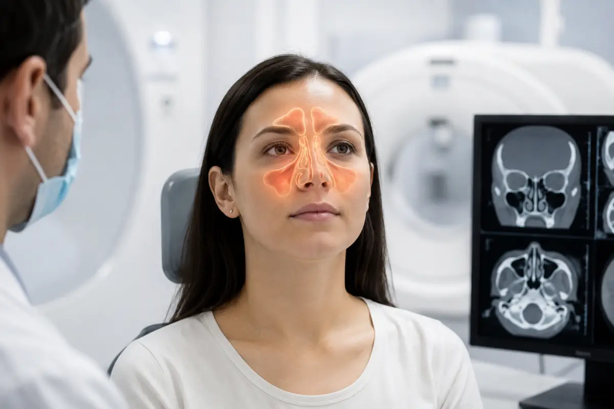
A CT scan of the paranasal sinuses is an X-ray examination that provides cross-sectional images of the nasal cavity, paranasal sinuses, and surrounding bony structures. The examination helps assess sinus drainage, identify signs of inflammation, cysts, polyps, anatomical variations, the consequences of trauma, and other changes.
At HBMedical, you can undergo a CT scan of the sinuses in Kyiv using a Siemens SOMATOM go.Up computed tomography scanner. The system performs 32 slices and up to 64 slices in IVR mode and also supports low-dose examinations. The center is located at 3 Oleksandra Dovzhenka Street, Kyiv, near «Shuliavska» metro station.
A standard CT scan of the paranasal sinuses is usually performed without intravenous contrast. Contrast enhancement may be prescribed for specific indications, for example, when a tumor, spread of infection, or complications involving the orbits and adjacent tissues are suspected.
You can schedule a CT scan of the paranasal sinuses at HBMedical by phone or through the online form. When booking, provide the exact wording from the referral: CT of the paranasal sinuses, CT of the nose and sinuses, or CT of the facial skeleton with 3D reconstruction.
Price of a CT scan of the paranasal sinuses in Kyiv
According to the current HBMedical price list, the examination costs:
- CT of the paranasal sinuses without contrast — UAH 2,100;
- CT of the paranasal sinuses with contrast enhancement — UAH 3,800;
- CT of the facial bones and paranasal sinuses with 3D reconstruction without contrast — UAH 2,350;
- CT of the facial bones and paranasal sinuses with 3D reconstruction and contrast enhancement — UAH 4,050.
The examination price includes a radiologist’s report and a copy of the results on a USB flash drive. Before your visit, it is recommended to confirm the current price and the required protocol with the administrator.
For diagnosing most inflammatory sinus conditions, a non-contrast CT scan is usually sufficient. There is no need to choose the more expensive option yourself: the need for contrast is determined by the doctor based on the symptoms and the clinical objective.
CT of the paranasal sinuses — what kind of examination is it?
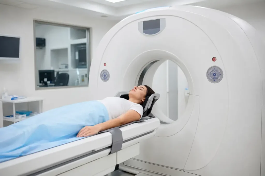
CT of the paranasal sinuses is computed tomography of the air-filled cavities surrounding the nasal cavity. The term «sinus CT» is also commonly used in medical practice.
The paranasal sinuses include:
- the maxillary sinuses;
- the frontal sinuses;
- the ethmoid air cells;
- the sphenoid sinus.
These cavities are located in the bones of the facial skeleton and communicate with the nasal cavity through small openings. If mucus drainage is impaired, swelling, inflammation, and fluid accumulation may develop in the sinuses.
During CT, the X-ray system creates a series of thin cross-sectional images. The doctor can review them in different planes and evaluate both the bony structures and the contents of the sinuses. CT is considered one of the most informative methods for assessing the anatomy and drainage pathways of the paranasal sinuses.
What is included in a sinus CT scan?
During a standard CT scan of the paranasal sinuses, the doctor evaluates:
- the maxillary sinuses;
- the frontal sinuses;
- the sphenoid sinus;
- the ethmoid labyrinth;
- the nasal cavity;
- the nasal septum;
- the nasal turbinates;
- the natural sinus openings;
- the ostiomeatal complexes;
- the bony sinus walls;
- adjacent parts of the facial skeleton;
- partially visualized orbital structures;
- the relationship between the upper teeth and the maxillary sinuses.
The scanned area depends on the selected protocol. A standard sinus CT scan is not a complete examination of the brain, inner ear, maxillofacial region, or soft tissues of the neck.
What does a CT scan of the paranasal sinuses show?
Computed tomography helps assess the condition of the sinuses and the causes of impaired ventilation and drainage.
A CT scan may show:
- thickening of the mucous membrane;
- accumulation of fluid or purulent contents;
- an air-fluid level in a sinus;
- partial or complete opacification of a sinus;
- acute or chronic sinusitis;
- maxillary sinusitis;
- frontal sinusitis;
- ethmoiditis;
- sphenoiditis;
- cysts and polypoid lesions;
- chronic rhinosinusitis with nasal polyps;
- obstruction of the natural sinus openings;
- narrowing of the ostiomeatal complexes;
- deviation of the nasal septum;
- enlargement or anatomical variations of the nasal turbinates;
- congenital anatomical variations of the sinuses;
- fungal involvement;
- foreign bodies;
- odontogenic inflammation associated with the upper teeth;
- fractures of the nasal bones and facial skeleton;
- postoperative changes;
- suspicious lesions in the nasal cavity or sinuses.
CT clearly shows bony anatomy and the presence of material within the sinuses. If the spread of a lesion into the soft tissues, brain, or orbits needs to be assessed in greater detail, the doctor may additionally recommend MRI.
Sinus CT for maxillary sinusitis and sinusitis
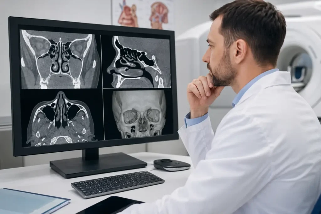
CT for maxillary sinusitis helps assess the maxillary sinuses, mucosal thickness, the presence of fluid, cysts, or polyps, and the patency of the natural openings.
The examination may be prescribed if:
- sinusitis symptoms persist or recur;
- treatment has not produced the expected result;
- frequent exacerbations occur;
- chronic rhinosinusitis is suspected;
- the doctor is planning surgery;
- the anatomical cause of impaired drainage needs to be clarified;
- odontogenic maxillary sinusitis is suspected;
- there are signs that inflammation has spread to adjacent structures.
CT is not always necessary for an uncomplicated acute runny nose or a first episode of acute sinusitis. The ENT specialist determines whether the examination is appropriate based on the symptoms, duration of illness, and examination findings. For chronic or recurrent rhinosinusitis and surgical planning, non-contrast CT is often the primary imaging method.
Indications for CT of the paranasal sinuses
A doctor may recommend a CT scan of the paranasal sinuses for the following symptoms and conditions:
- persistent nasal congestion;
- recurrent or chronic sinusitis;
- pain or pressure in the face;
- pain over the maxillary or frontal sinuses;
- purulent nasal discharge;
- reduced or lost sense of smell;
- headache accompanied by ENT symptoms;
- suspected cyst or polyp;
- one-sided symptoms;
- frequent nosebleeds;
- suspected fungal sinusitis;
- trauma to the nose or facial skeleton;
- suspected foreign body;
- changes detected during rhinoscopy;
- preparation for endoscopic sinus surgery;
- follow-up after surgical treatment;
- the need to clarify the findings of radiography, ultrasound, or another examination.
One-sided sinus opacification, bloody discharge, or gradually worsening symptoms require medical evaluation. These signs do not necessarily indicate a tumor, but they may require an extended diagnostic work-up.
Sinus CT with contrast
Intravenous contrast is usually not required for a standard sinus examination in patients with sinusitis. Non-contrast CT clearly shows bony anatomy, mucosal thickening, fluid, polyps, cysts, and obstruction of the drainage pathways.
Contrast-enhanced CT of the paranasal sinuses may be recommended for:
- suspected tumor of the nasal cavity or sinuses;
- the need to assess the extent of a lesion;
- suspected abscess;
- possible spread of infection into the orbit;
- suspected intracranial complications;
- complex postoperative changes;
- the need to distinguish inflammatory tissue from another type of lesion.
If a contrast-enhanced CT scan is planned, inform the staff about:
- kidney disease;
- thyroid disease;
- previous reactions to iodine-containing contrast agents;
- bronchial asthma or severe allergic reactions;
- diabetes and any medications you take;
- pregnancy or the possibility of pregnancy.
If risk factors are present, a blood creatinine test may be required before contrast administration to assess kidney function.
3D CT of the sinuses
CT images can be reconstructed in different planes and used to create three-dimensional models. 3D reconstruction is particularly useful for assessing bony structures, complex anatomical relationships, trauma, and surgical planning.
HBMedical also offers a dedicated CT protocol of the facial bones and paranasal sinuses with 3D reconstruction. This protocol may be recommended for:
- facial bone injuries;
- fractures;
- complex anatomical deformities;
- preparation for maxillofacial surgery;
- planning sinus surgery;
- the need to evaluate the sinuses and facial bones at the same time.
A separate extended 3D protocol is not necessary for every patient undergoing routine evaluation for sinusitis.
CT of the nose and nasopharynx: is it the same examination?
A sinus CT scan shows the nasal cavity, paranasal sinuses, and surrounding bony structures. Part of the nasopharynx may be included in the scanning area, but this does not mean that a standard sinus CT fully replaces a targeted examination of the nasopharynx.
If the doctor needs to assess:
- the soft tissues of the nasopharynx;
- the larynx or throat;
- the cervical lymph nodes;
- the spread of a tumor;
- the deep spaces of the neck,
a contrast-enhanced CT scan of the neck soft tissues or MRI may be required. When booking, provide the exact wording from the referral to avoid undergoing the wrong protocol.
Sinus CT and facial bone CT: what is the difference?
CT of the paranasal sinuses
The main focus is on:
- the condition of the sinuses;
- the mucous membrane;
- the natural openings;
- the nasal septum;
- anatomy relevant to the ENT specialist.
CT of the facial bones
This examination is more focused on assessing:
- the nasal bones;
- the zygomatic bones;
- the upper and lower jaws;
- the orbital walls;
- fractures and deformities;
- the consequences of trauma.
CT of the facial skeleton and sinuses with 3D reconstruction
This protocol combines assessment of the sinuses and facial bony structures. It may be needed after trauma or before surgery.
CT or X-ray of the sinuses
Radiography creates a two-dimensional image in which anatomical structures may overlap. It can show marked opacification or an air-fluid level, but it provides less detailed visualization of individual sinuses, natural openings, and complex anatomy.
CT produces thin cross-sectional images and helps assess all sinus groups, the nasal septum, ostiomeatal complexes, and bony walls in detail. Therefore, computed tomography is generally more informative for chronic or recurrent rhinosinusitis and surgical planning.
Do not replace one method with another on your own. The examination should correspond to the doctor’s clinical objective.
How to prepare for a sinus CT scan
No special preparation is usually required for a non-contrast CT scan of the paranasal sinuses.
Before the examination, it is recommended to:
- bring a referral from an ENT specialist or another doctor;
- bring previous CT, MRI, or X-ray images;
- bring discharge summaries after surgery on the nose or sinuses;
- inform the staff about pregnancy or the possibility of pregnancy;
- remove earrings, piercings, glasses, hearing aids, and metal jewelry;
- inform the staff about any non-removable metal structures in the facial area;
- bring previous examination results for comparison.
Dental implants, fillings, or metal structures usually do not prevent CT scanning, but they may create artifacts on the images.
Preparation for contrast-enhanced CT
If the doctor has prescribed contrast enhancement, clarify the following when booking:
- how many hours before the procedure you may eat;
- whether a creatinine test is required;
- which documents and test results you should bring;
- whether the schedule for taking certain medications needs to be changed.
Do not stop taking any medication on your own. Decisions about medication are made by the doctor.
How is a sinus CT scan performed?
Before the examination begins, the patient leaves metal items outside the scanning room and completes a questionnaire if required.
The procedure takes place in several stages:
- Positioning. The patient lies down on the movable table of the CT scanner.
- Head stabilization. The head is positioned correctly and may be secured with soft supports to prevent movement.
- Scanning. The table moves through the scanner ring while the system creates a series of cross-sectional images of the nose and sinuses.
- Contrast administration. If contrast-enhanced CT has been prescribed, the contrast agent is administered intravenously, after which the required series of images is obtained.
- Result processing. The radiologist reviews the images in different planes and prepares a report.
The scan itself takes only a short time. The procedure is painless, but it is important to keep the head still to obtain clear images.
Interpretation of a sinus CT scan
After the examination, the patient receives a radiologist’s report. It may contain specialized terms.
The sinuses are aerated
This means that the sinuses are filled with air. Preserved aeration without fluid or marked mucosal thickening usually corresponds to a normal appearance.
Mucosal thickening
This means that the lining of the sinus has become thicker. This change may be seen with inflammation, allergy, or after a recent infection. Mild thickening does not always explain the symptoms and may be an incidental finding.
Air-fluid level
A horizontal fluid level may be seen in acute inflammation, but it must be interpreted together with the symptoms and examination findings.
Partial or complete sinus opacification
This means that the sinus is partly or completely filled with mucus, fluid, thickened mucosa, or other material instead of air.
Maxillary sinus cyst
A retention cyst is a rounded lesion associated with the mucous membrane. Not every cyst requires treatment. Its significance is determined by the ENT specialist based on its size, location, and the patient’s symptoms.
Polypoid mucosal thickening
This may indicate localized mucosal overgrowth or polypoid changes. Establishing a diagnosis requires correlation with an ENT examination.
The ostiomeatal complex is obstructed
This means that the main drainage pathway from the frontal sinus, maxillary sinus, and anterior ethmoid air cells is narrowed or blocked.
Deviated nasal septum
CT shows the direction and severity of the deviation. However, the need for treatment depends not only on the image but also on impaired nasal breathing and examination findings.
Do not try to determine a diagnosis from a single phrase in the report. The CT results should be reviewed by an ENT specialist.
What does a normal CT scan of the paranasal sinuses look like?
On a normal CT scan, the paranasal sinuses are generally:
- filled with air;
- free of air-fluid levels;
- without marked mucosal thickening;
- surrounded by intact bony walls;
- connected to the nasal cavity through patent openings.
At the same time, anatomical variants, slight asymmetry, or minimal mucosal thickening may also be present in people without significant symptoms. A radiologist determines whether the scan is normal after reviewing all slices.
Contraindications and limitations
CT uses X-rays, so the examination is performed for medical indications.
Before the procedure, inform the staff about:
- pregnancy or the possibility of pregnancy;
- recent CT examinations;
- an inability to remain still;
- severe pain or other conditions that make scanning difficult.
CT is generally not performed during pregnancy unless there is an urgent medical need. The final decision is made by the doctor after assessing the expected benefit and potential risk.
Pacemakers, orthopedic implants, and most metal devices are not contraindications to CT. However, metal within the scanned area may reduce image quality.
What does the patient receive after CT?
After the examination, the radiologist reviews the images and prepares a report.
The patient receives:
- CT images of the paranasal sinuses;
- a radiologist’s report;
- a copy of the examination on a USB flash drive;
- results for consultation with an ENT specialist, maxillofacial surgeon, dentist, or another specialist.
At HBMedical, CT results are usually prepared within 5–7 hours. In complex cases, the timeframe may vary. The report describes the detected changes, but the final diagnosis is made by the relevant specialist.
Why choose HBMedical for a sinus CT scan?
- Siemens SOMATOM go.Up computed tomography scanner;
- 32 slices and up to 64 slices in IVR mode;
- the option of a low-dose examination;
- CT of the paranasal sinuses without and with contrast enhancement;
- a dedicated facial skeleton and sinus protocol with 3D reconstruction;
- image interpretation by a radiologist;
- the report and USB flash drive are included in the price;
- the option to compare findings with previous examinations;
- the diagnostic department is open daily;
- convenient location near «Shuliavska» metro station;
- advance online booking.
Where to get a sinus CT scan in Kyiv
You can undergo a CT scan of the paranasal sinuses at HBMedical, located at 3 Oleksandra Dovzhenka Street, Kyiv, near «Shuliavska» metro station.
The center is located on the right bank of Kyiv. It is convenient for patients coming from Shuliavka, KPI, Lukianivka, Dorohozhychi, Nyvky, as well as the Shevchenkivskyi, Solomianskyi, and Sviatoshynskyi districts.
When booking, inform the administrator about:
- the exact name of the examination stated in the referral;
- whether contrast enhancement is required;
- whether only the sinuses or also the facial bones need to be examined;
- whether surgery is planned;
- whether previous CT or MRI scans are available;
- whether you have undergone surgery on the nose or sinuses;
- whether you have kidney disease or previous reactions to contrast agents;
- whether you are pregnant or may be pregnant.
Schedule a CT scan of the paranasal sinuses at HBMedical to undergo the examination in Kyiv near Shuliavska and receive a report for further consultation with an ENT specialist.
Frequently asked questions
What is a CT scan of the paranasal sinuses?
It is computed tomography of the air-filled sinuses around the nasal cavity. The examination shows the maxillary, frontal, and sphenoid sinuses, the ethmoid air cells, the nasal cavity, and the drainage pathways.
What does a sinus CT scan show?
The examination may show sinusitis, maxillary sinusitis, mucosal thickening, fluid, cysts, polyps, fungal changes, a deviated septum, obstruction of the natural openings, and the consequences of trauma.
How much does a sinus CT scan cost in Kyiv?
According to the current HBMedical price list, a non-contrast CT scan of the paranasal sinuses costs UAH 2,100, while a contrast-enhanced scan costs UAH 3,800. CT of the facial skeleton and sinuses with 3D reconstruction costs UAH 2,350 without contrast and UAH 4,050 with contrast.
Is contrast needed for a sinus CT scan?
Contrast is not required for diagnosing most cases of sinusitis. It may be prescribed if a tumor, abscess, spread of infection, or other complications are suspected.
How should I prepare for a sinus CT scan?
No special preparation is usually required for a non-contrast examination. Metal items in the head area should be removed, and the staff should be informed about a possible pregnancy.
How is a CT scan of the paranasal sinuses performed?
The patient lies on the scanner table while the system creates a series of cross-sectional images of the nose and sinuses. The head must remain still during scanning.
Can maxillary sinusitis be seen on CT?
Yes. CT may show thickening of the maxillary sinus mucosa, fluid, complete or partial opacification, a cyst, a polyp, and impaired drainage.
Can a sinus CT scan be performed when I have a runny nose?
Yes, but a routine acute runny nose is not always an indication for CT. The doctor determines whether the examination is necessary based on the duration and severity of the symptoms.
Is the nasopharynx included in a sinus CT scan?
Part of the nasopharynx may be visible, but a standard sinus CT does not replace a targeted examination of the nasopharyngeal or neck soft tissues.
Are sinus CT and 3D CT the same examination?
Three-dimensional reconstructions can be created from CT images. HBMedical offers a separate extended CT protocol of the facial bones and paranasal sinuses with 3D reconstruction.
What does mucosal thickening on CT mean?
It means that the lining of the sinus has increased in thickness. It may be associated with inflammation, allergy, or a recent infection and is interpreted together with the symptoms.
When will the report be ready?
At HBMedical, CT results are usually prepared within 5–7 hours. In complex cases, preparing the report may take longer.
This material is for informational purposes only and does not replace a medical consultation.










Мені все сподобалось
Всі профі
Дякую


Велика подяка лікарю Дуброву Є.О. — надзвичайно компетентний і чуйний фахівець, який детально все пояснив, розвіяв тривогу та підтримав морально. Щиро дякую анестезіологу Бєлаш О.М. за високий професіоналізм, чуйність і справжнє людське ставлення — занурення в сон та сам перебіг процедури минули максимально комфортно. Також величезне спасибі медсестрі (на жаль, не дізналася ім'я) за її турботу, доброту та увагу.
Весь персонал вразив своєю чуйністю та теплом. Дякую за ваш труд і турботу про пацієнтів! Однозначно рекомендую.














Дуже ввічливий та приємний персонал клініки. Лікар був уважний, без поспіху пройшовся по всім аспектам здоровʼя. Лабораторія швидко зробила аналізи. В цілому дуже задоволена. Радже звертатися!




Саме до таких фахівців, в разі необхідності, є бажання повернутися!

і закінчуючи отриманим описом і рекомендаціями від лікарів.
























Need specialist help?
Order a callback and our consultant will help you choose a doctor and make an appointment at a convenient time for you!