








 Work time
Diagnostics: Mon - Sun, 6:00 - 23:00
Polyclinic: Mon - Sun, 8:00 - 20:00

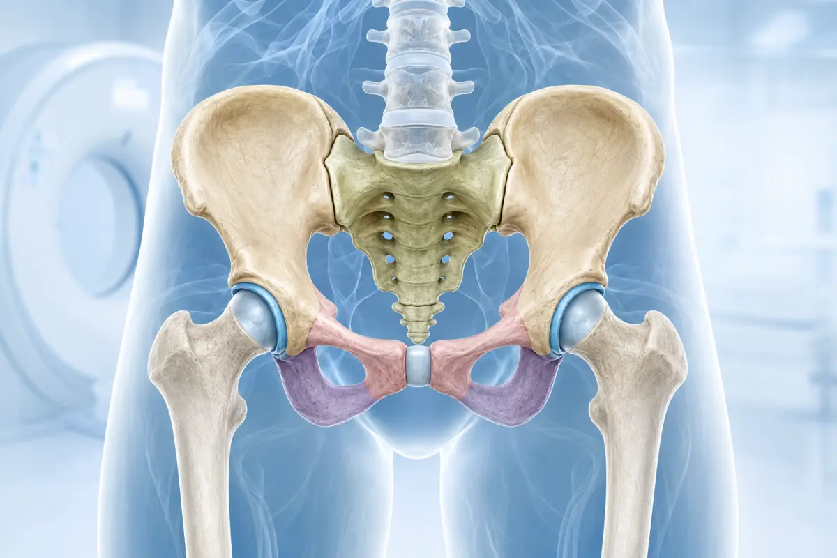
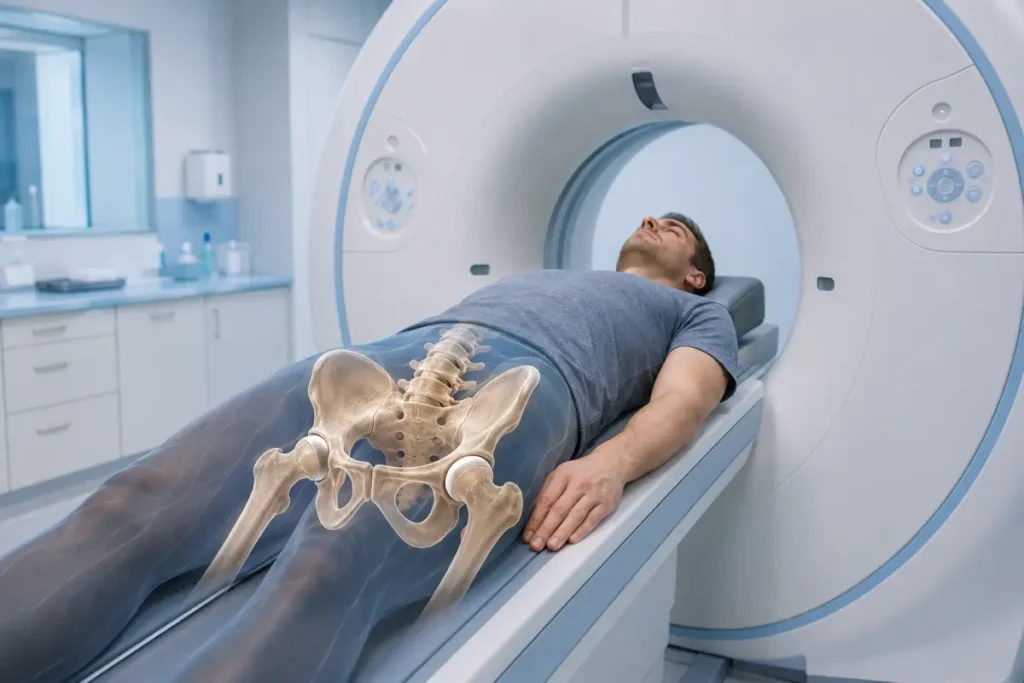
A pelvic bone CT is a cross-sectional X-ray examination that helps assess the pelvic ring, acetabula, sacroiliac joints, pubic symphysis and adjacent bone structures in detail. Computed tomography is used to detect fractures, cracks, displacement of bone fragments, deformities, neoplasms, inflammatory changes and postoperative findings.
At the HBMedical medical center, you can undergo a pelvic bone CT in Kyiv as well as a comprehensive CT scan of the pelvic bones and hip joints. The examination is performed using a Siemens SOMATOM go.Up CT scanner with 32 slices and up to 64 slices in IVR mode.
Center address: 3 Oleksandra Dovzhenka Street, Kyiv, near the «Shuliavska» metro station.
For the assessment of fractures and other structural bone changes, a non-contrast CT scan is usually sufficient. A contrast agent may be required if a tumor, inflammation, abscess or spread of a pathological process into the surrounding soft tissues is suspected.
You can schedule a pelvic bone CT at HBMedical by phone or through the form on the website. When booking, provide the exact wording from the referral: pelvic bone CT, hip joint CT or a comprehensive examination of the pelvic bones and hip joints.
Pelvic Bone CT Price in Kyiv
According to the current HBMedical price list, the following examinations are available:
- pelvic bone CT without contrast — UAH 1,850;
- pelvic bone CT with contrast enhancement — UAH 3,300;
- pelvic bone and hip joint CT without contrast — UAH 2,500;
- pelvic bone and hip joint CT with contrast enhancement — UAH 2,600.
The examination fee includes a radiologist’s report and a USB flash drive containing the results. Before your visit, it is advisable to confirm the current price and the exact scope of the service, especially if the doctor has prescribed contrast enhancement or a comprehensive examination of the hip joints.
The required protocol depends on the location of the pain and the diagnostic objective. A pelvic bone CT may be sufficient for an injury to the pelvic ring. If the femoral heads, acetabula and both hip joints need to be assessed in detail, a comprehensive examination should be booked.
What Is a Pelvic Bone CT?
The pelvis is a complex bony structure that connects the spine to the lower limbs, supports the pelvic organs and helps distribute load while walking.
The bony pelvic ring is formed by:
- the iliac bones;
- the ischial bones;
- the pubic bones;
- the sacrum;
- the pubic symphysis;
- the right and left sacroiliac joints.
The acetabula are located on both sides of the pelvis and contain the heads of the femurs. Together, these structures form the hip joints.
Because of the complex anatomy of the pelvis, standard radiography does not always show every fracture line or the degree of bone fragment displacement. CT creates thin cross-sectional images that can be viewed in different planes and used to generate three-dimensional reconstructions.
What Is Included in a Pelvic Bone CT?
Within a standard protocol, the radiologist may assess:
- the iliac bones;
- the ischial bones;
- the pubic bones;
- the iliac wings;
- the superior and inferior pubic rami;
- the ischial tuberosities;
- the acetabula;
- the sacroiliac joints;
- the pubic symphysis;
- the sacrum within the scan area;
- bone canals and foramina;
- the integrity of the pelvic ring;
- adjacent portions of the femurs;
- surrounding soft tissues within the capabilities of CT.
The exact scan boundaries depend on the protocol and the doctor’s referral. If the main symptoms are localized in the hip joint, sacrum, coccyx or pelvic organs, a separate or extended examination may be required.
What Does a Pelvic Bone CT Show?
CT helps assess the integrity, density, shape and relative position of the bones in the pelvic region in detail.
The examination may show:
- pelvic bone fractures;
- bone cracks and occult fractures;
- acetabular fractures;
- sacral injuries;
- disruption of the pelvic ring;
- displacement of bone fragments;
- bone impaction or deformity;
- hip dislocation or subluxation;
- separation of the pubic symphysis;
- injury to the sacroiliac joints;
- consequences of an old injury;
- malunited fractures;
- degenerative changes;
- bone spurs;
- areas of bone destruction or remodeling;
- benign and suspicious bone lesions;
- signs of inflammatory bone involvement;
- postoperative changes;
- the position of plates, screws and other fixation devices;
- the condition of bone tissue around implants;
- congenital anatomical features of the pelvis.
CT is particularly informative for evaluating bone structures. In many clinical situations, ligaments, tendons, the acetabular labrum, muscles and bone marrow are better assessed with MRI.
Pelvic Bone CT After an Injury
A pelvic bone injury may result from:
- a road traffic accident;
- a fall from height;
- a strong impact;
- a sports injury;
- an occupational accident;
- a fall onto the side or buttocks;
- osteoporosis combined with a minor injury in older age.
CT may be prescribed if the following symptoms occur after an injury:
- severe pelvic pain;
- pain in the groin, buttocks or hip region;
- inability to bear weight on the leg;
- severe restriction of movement;
- deformity of the pelvic region;
- significant swelling or hematoma;
- asymmetry in the length or position of the lower limbs;
- pain when the pelvis is compressed;
- urination problems;
- blood in the urine;
- neurological symptoms in the lower limbs;
- suspected fracture following an inconclusive X-ray result.
In the event of a serious injury, the examination is performed as part of emergency care. If simultaneous damage to internal organs or blood vessels is suspected, the doctor may order not only a bone CT but also a comprehensive contrast-enhanced CT scan of the abdomen and pelvis.
CT makes it possible to determine the extent of bone injury more accurately than a standard X-ray and is used to assess complex fractures and plan surgical treatment.
Which Pelvic Fractures Can Be Seen on CT?
Computed tomography helps detect and characterize:
- fractures of the iliac wing;
- fractures of the superior and inferior pubic rami;
- fractures of the ischium;
- sacral fractures;
- acetabular fractures;
- marginal pelvic fractures;
- multiple fractures of the pelvic ring;
- compression and impacted injuries;
- stress and insufficiency fractures;
- old fractures and impaired fracture healing.
In the report, the doctor may describe the location of the fracture line, the degree of bone fragment displacement and the involvement of the articular surfaces, pubic symphysis or sacroiliac joints.
CT of the Pelvic Bones and Hip Joints
CT of the pelvic bones and hip joints is an extended protocol that allows the pelvic ring and both hip joints to be assessed at the same time.
During a comprehensive examination, the following may be evaluated:
- the right and left acetabula;
- the femoral heads;
- the femoral necks;
- the proximal portions of the femurs;
- the shape and alignment of the articular surfaces;
- the width of the joint spaces;
- bone spurs;
- subchondral sclerosis;
- cystic changes;
- fractures and deformities;
- dislocations and subluxations;
- the position of hip replacements or other implants;
- the condition of the bone tissue around an implant.
A hip joint CT may be prescribed after an injury, in cases of osteoarthritis or complex deformity, before joint replacement surgery or for postoperative follow-up.
If a fracture in the hip region is suspected after an inconclusive X-ray, a non-contrast CT may be used as the next imaging method.
Pelvic Bone CT and CT of the Pelvic Organs — What Is the Difference?
These are different examinations with different diagnostic objectives.
Pelvic bone CT
The main focus is on:
- the pelvic ring;
- the acetabula;
- the sacroiliac joints;
- the pubic symphysis;
- fractures;
- bone lesions;
- deformities;
- postoperative changes.
CT of the pelvic organs
This examination is intended to assess:
- the urinary bladder;
- the rectum;
- the uterus and adnexa in women;
- the prostate gland and seminal vesicles in men;
- the lymph nodes;
- the soft tissues and pelvic fat.
The bones are also visible during a CT scan of the pelvic organs, but the protocol and primary clinical objective are different. If the doctor has prescribed a pelvic bone CT, it should not be replaced with a CT scan of the pelvic organs without medical advice.
Pelvic CT and Hip Joint CT — What Is the Difference?
A pelvic bone CT covers a broad area of the pelvic ring. A separate CT scan of one hip joint has a smaller scan area and may provide a more detailed targeted assessment of that particular joint.
When booking, clarify:
- whether the pain is on one side or both sides;
- whether there has been an injury;
- whether the entire pelvis needs to be assessed;
- whether a joint replacement is present;
- whether surgery is being planned;
- whether the referral specifies one or both hip joints.
At HBMedical, pelvic bone CT, CT of the pelvic bones and hip joints, and CT of a single joint are listed as separate services.
Contrast-Enhanced Pelvic Bone CT
Intravenous contrast is not required to diagnose most fractures and structural bone changes. Bone tissue is clearly visualized on a standard non-contrast scan.
A contrast-enhanced pelvic bone CT may be recommended when it is necessary to assess:
- a tumor lesion;
- the spread of a pathological process into the soft tissues;
- an inflammatory infiltrate;
- an abscess;
- osteomyelitis;
- postoperative complications;
- blood vessels;
- internal bleeding after an injury;
- the condition of adjacent organs and tissues.
In a severe pelvic injury, contrast-enhanced CT may be used not only to assess the bones but also to detect damage to blood vessels, the urinary bladder, the intestines and other organs. In this case, a different, extended protocol is performed.
Contrast enhancement should not be selected independently. The doctor determines whether a contrast agent is required based on the clinical objective.
When Is a Pelvic Bone CT Prescribed?
A referral for the examination may be issued by an orthopedic traumatologist, surgeon, oncologist, rheumatologist or another doctor.
Main indications include:
- pelvic injury;
- suspected fracture;
- an inconclusive X-ray result;
- pelvic pain of unclear origin;
- inability to bear weight fully on the leg;
- deformity of the pelvic ring;
- acetabular injury;
- pain in the sacroiliac region;
- suspected bone tumor;
- a detected area of bone destruction;
- suspected osteomyelitis;
- congenital deformity;
- preparation for surgery;
- planning reconstructive treatment;
- preparation for joint replacement surgery;
- assessment of fracture healing;
- postoperative follow-up;
- assessment of the position of plates, screws or a prosthesis;
- clarification of X-ray, MRI or other examination findings.
Pelvic Bone CT for Tumors
CT can help assess a bone lesion and determine:
- its location;
- size;
- the definition of its margins;
- structure;
- density;
- the presence of calcifications;
- destruction of the cortical bone;
- the reaction of the surrounding bone tissue;
- a pathological fracture;
- spread into adjacent structures.
However, CT does not always make it possible to determine the exact type of lesion. The doctor may additionally prescribe MRI to assess the bone marrow and soft-tissue component. A definitive diagnosis may require a biopsy and histological examination.
CT of the Sacroiliac Joints
The sacroiliac joints connect the sacrum and the iliac bones. They are part of the pelvic region and can be assessed during a pelvic bone CT.
The examination may show:
- narrowing or irregularity of the joint space;
- subchondral sclerosis;
- erosions;
- bone spurs;
- ankylosis;
- consequences of injury;
- a fracture;
- postoperative changes.
If active inflammation of the sacroiliac joints is suspected, the doctor may recommend MRI because it is better at showing bone marrow edema and early inflammatory changes. CT is primarily useful for assessing structural bone changes.
Pelvic CT in Osteoporosis
Osteoporosis increases the risk of fractures even after a minor fall. In older adults, fractures of the sacrum, pubic bones or other pelvic bones may be difficult to see on initial X-rays.
CT can help detect an occult fracture and determine its location. However, pelvic bone CT itself is not a standard method for diagnosing osteoporosis. DXA densitometry is most commonly used to assess bone mineral density.
Pelvic Bone CT or MRI
CT and MRI have different diagnostic capabilities.
CT is better at showing:
- fractures;
- bone cracks;
- displacement of bone fragments;
- the cortical layer of bone;
- calcifications;
- bone deformities;
- the position of metal fixation devices;
- the condition of the acetabula;
- the complex three-dimensional anatomy of the pelvis.
MRI is better suited for evaluating:
- bone marrow;
- occult bone edema;
- muscles;
- tendons and ligaments;
- the acetabular labrum;
- cartilage;
- nerves;
- early inflammatory changes;
- the soft-tissue component of a tumor.
The doctor may prescribe both examinations when a comprehensive assessment of bone and soft-tissue changes is required.
Pelvic Bone CT or X-Ray
Radiography is often the first examination used for pelvic pain or injury. It provides a general image of the bones, but the overlap of anatomical structures may conceal certain fractures.
CT produces cross-sectional images and helps:
- clarify the fracture line;
- identify additional injuries;
- assess the acetabulum;
- determine the displacement of bone fragments;
- evaluate the posterior portions of the pelvic ring;
- prepare for surgery.
Do not replace radiography with computed tomography on your own. The doctor determines the appropriate diagnostic sequence.
Preparation for a Pelvic Bone CT
Special preparation is usually not required for a non-contrast pelvic bone CT.
Before the examination, it is recommended to:
- bring the doctor’s referral;
- bring previous X-rays, CT scans or MRI scans;
- bring medical records following an injury or surgery;
- report any prostheses, plates or screws;
- report a possible pregnancy;
- remove metal objects from the scan area;
- wear comfortable clothing without metal fittings;
- tell the staff if pain prevents you from lying still;
- do not stop regular medication without a doctor’s advice.
A metal joint replacement, plate or screws are not an absolute obstacle to CT. However, metal may create artifacts and partially reduce image quality.
Preparation for a Contrast-Enhanced Pelvic Bone CT
If intravenous contrast enhancement has been prescribed, inform the staff about:
- kidney disease;
- reduced kidney function;
- previous reactions to iodine-containing contrast agents;
- thyroid disease;
- bronchial asthma;
- diabetes mellitus;
- severe allergic reactions;
- the medications you are taking;
- pregnancy or the possibility of pregnancy.
An up-to-date blood creatinine test may be required before a contrast-enhanced CT scan. Dietary instructions, fluid intake requirements and the acceptable age of the test result should be clarified when booking.
Do not stop taking medication on your own. Recommendations regarding medication should be provided by a doctor or the diagnostic center staff.
How Is a Pelvic Bone CT Performed?
Before the procedure, the patient leaves metal objects outside the examination room and completes a questionnaire if necessary.
The examination is performed in several stages:
- Positioning. The patient lies on the movable CT table, usually on their back.
- Position stabilization. The body is positioned so that the pelvic region is centered in the scan area. Soft supports may be used if necessary.
- Scanning. The table moves through the scanner ring while the system creates a series of thin cross-sectional images.
- Contrast enhancement. If a contrast-enhanced examination has been prescribed, the agent is administered intravenously and an additional image series is acquired.
- Reconstruction. The images are processed in the coronal, sagittal and other planes. Three-dimensional images may also be generated when required.
- Report preparation. The radiologist analyzes the images and prepares a written report.
The procedure is painless. To obtain clear images, it is important to remain still and follow the medical staff’s instructions.
How Long Does a Pelvic Bone CT Take?
The scan itself is usually brief. The total time spent in the diagnostic department depends on:
- the time required to complete documents;
- the complexity of positioning the patient after an injury;
- the selected protocol;
- intravenous contrast enhancement;
- the need for additional reconstructions.
It is advisable to ask for an approximate duration when booking.
Contraindications and Limitations
CT uses ionizing radiation, so the examination is performed when medically indicated.
Before the procedure, inform the staff about:
- pregnancy or the possibility of pregnancy;
- recent CT examinations;
- an inability to remain still;
- severe pain;
- a condition that makes correct body positioning difficult;
- the need for assistance during transfers.
During pregnancy, CT of the pelvic region is generally avoided unless there is an urgent need. In an emergency, the doctor makes the decision after assessing the expected benefit and potential risk.
For a contrast-enhanced examination, kidney function, previous reactions to iodine-containing agents and certain thyroid conditions are also taken into account.
What Does the Patient Receive After the CT Scan?
After scanning, the radiologist analyzes the cross-sectional images and reconstructions.
The patient receives:
- CT images of the pelvic bones;
- a radiologist’s report;
- the examination recorded on a USB flash drive;
- materials for consultation with an orthopedic traumatologist, surgeon, oncologist or another specialist.
At HBMedical, CT results are usually prepared within 5–7 hours. In complex cases, the turnaround time may vary. The report describes the detected changes, but the final diagnosis and treatment plan are determined by the relevant specialist.
Why Choose HBMedical for a Pelvic Bone CT?
- Siemens SOMATOM go.Up CT scanner;
- 32 slices and up to 64 slices in IVR mode;
- the option to use low-dose protocols;
- pelvic bone CT with and without contrast;
- comprehensive CT of the pelvis and hip joints;
- detailed assessment of fractures and bone deformities;
- the option to generate multiplanar reconstructions;
- image interpretation by a radiologist;
- the report and USB flash drive are included in the price;
- the option to compare the findings with previous examinations;
- results are usually prepared within 5–7 hours;
- location near the «Shuliavska» metro station;
- advance booking by phone or online.
Where to Get a Pelvic Bone CT in Kyiv
You can undergo a pelvic bone CT at the HBMedical medical center at 3 Oleksandra Dovzhenka Street, Kyiv, near the «Shuliavska» metro station.
The center is located on the right bank of Kyiv. It is convenient for patients from Shuliavka, KPI, Lukianivka, Dorohozhychi and Nyvky, as well as the Shevchenkivskyi, Solomianskyi and Sviatoshynskyi districts.
When booking, tell the administrator:
- the exact name of the examination stated in the referral;
- whether there has been an injury;
- where the pain is located;
- whether the hip joints need to be assessed;
- whether contrast enhancement is required;
- whether previous X-rays, CT scans or MRI scans are available;
- whether any operations have been performed;
- whether prostheses or metal fixation devices are present;
- whether there is kidney disease or a history of reactions to contrast agents;
- whether there is a pregnancy or a possibility of pregnancy.
Schedule a pelvic bone CT at HBMedical to undergo the examination in Kyiv near Shuliavska and receive the results for further consultation with the relevant specialist.
Frequently Asked Questions
What does a pelvic bone CT show?
CT can help detect fractures, bone cracks, displacement of fragments, acetabular injuries, deformities, bone lesions, inflammatory changes and postoperative findings.
How much does a pelvic bone CT cost in Kyiv?
According to the current HBMedical price list, a non-contrast pelvic bone CT costs UAH 1,850, while a contrast-enhanced examination costs UAH 3,300.
What is the price of a CT scan of the pelvic bones and hip joints?
According to the HBMedical price list, a comprehensive non-contrast CT scan of the pelvic bones and hip joints costs UAH 2,500. It is advisable to confirm the price of the contrast-enhanced protocol when booking.
Are the hip joints included in a pelvic bone CT?
Parts of the hip joints may be included in the scan area, but a separate comprehensive CT protocol covering the pelvic bones and hip joints is preferable for a complete assessment.
Can an occult fracture be seen on CT?
CT can detect fractures that are difficult to see on standard radiographs. In some cases, especially when an insufficiency fracture or bone marrow injury is suspected, the doctor may also prescribe MRI.
Is contrast required for a pelvic fracture?
Contrast is not required to assess most fractures. In severe trauma, it may be used to detect internal bleeding and injuries to blood vessels, organs and soft tissues.
How should I prepare for a pelvic bone CT?
Special preparation is usually not required for a non-contrast examination. Bring the referral and previous images, and report a possible pregnancy.
How is a pelvic bone CT performed?
The patient lies on the movable CT table while the scanner creates a series of thin cross-sectional images of the pelvic region. It is important to remain still during scanning.
Can CT be performed with a metal plate or joint replacement?
Yes. A metal fixation device usually does not prevent a CT scan. However, metal can create artifacts, so it should be reported when booking.
How does a pelvic bone CT differ from a CT scan of the pelvic organs?
A pelvic bone CT is primarily intended to assess fractures and other changes in bone structures. A CT scan of the pelvic organs is used to examine the urinary bladder, intestines, reproductive organs, lymph nodes and soft tissues.
Which is better for the pelvic bones — CT or MRI?
CT is better at showing fractures, bone fragments and the cortical layer. MRI is more informative for assessing bone marrow, ligaments, muscles and early inflammatory changes.
Is a referral required?
You can ask whether the examination is available without a referral when booking. A referral is preferable because it helps select the correct examination among pelvic bone CT, hip joint CT and CT of the pelvic organs.
When will the report be ready?
At HBMedical, CT results are usually prepared within 5–7 hours. In complex cases, the turnaround time may be longer.
This material is for informational purposes only and does not replace a consultation with a doctor.










Мені все сподобалось
Всі профі
Дякую


Велика подяка лікарю Дуброву Є.О. — надзвичайно компетентний і чуйний фахівець, який детально все пояснив, розвіяв тривогу та підтримав морально. Щиро дякую анестезіологу Бєлаш О.М. за високий професіоналізм, чуйність і справжнє людське ставлення — занурення в сон та сам перебіг процедури минули максимально комфортно. Також величезне спасибі медсестрі (на жаль, не дізналася ім'я) за її турботу, доброту та увагу.
Весь персонал вразив своєю чуйністю та теплом. Дякую за ваш труд і турботу про пацієнтів! Однозначно рекомендую.














Дуже ввічливий та приємний персонал клініки. Лікар був уважний, без поспіху пройшовся по всім аспектам здоровʼя. Лабораторія швидко зробила аналізи. В цілому дуже задоволена. Радже звертатися!




Саме до таких фахівців, в разі необхідності, є бажання повернутися!

і закінчуючи отриманим описом і рекомендаціями від лікарів.
























Need specialist help?
Order a callback and our consultant will help you choose a doctor and make an appointment at a convenient time for you!