








 Work time
Diagnostics: Mon - Sun, 6:00 - 23:00
Polyclinic: Mon - Sun, 8:00 - 20:00

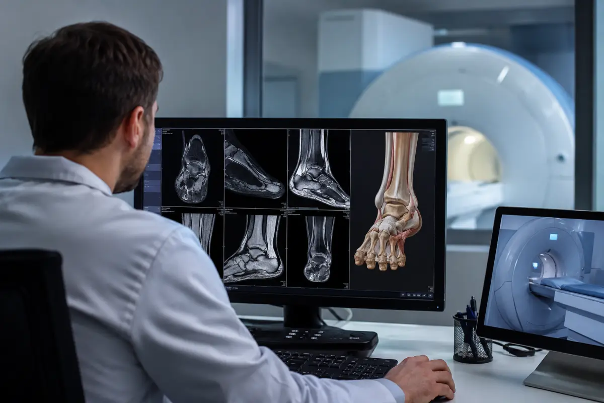
An ankle MRI is a detailed magnetic resonance examination of the bones, articular cartilage, ligaments, tendons and soft tissues in the ankle area. The examination helps determine the cause of pain, swelling, joint instability or limited mobility after an injury, physical activity or in the presence of chronic symptoms.
At the HBMedical medical center, you can undergo an MRI of the ankle joint and the adjacent proximal part of the foot. The examination is performed using a Siemens Magnetom Sempra 1.5 Tesla scanner at: 3 Oleksandra Dovzhenka Street, Kyiv, near Shuliavska metro station.
MRI does not use X-ray radiation and provides detailed images of the joints, bone marrow, cartilage, ligaments, tendons and surrounding soft tissues. The method is especially useful for detecting injuries that may not be visible on a standard X-ray.
You can book an ankle MRI at HBMedical by phone or through the online form. When booking, provide the exact area specified in the referral and indicate which leg needs to be examined.
Ankle MRI price in Kyiv
The following prices apply at HBMedical:
- MRI of the ankle joint and proximal foot without contrast — UAH 3,750;
- Ankle MRI with contrast enhancement — UAH 6,500.
The examination price includes a radiologist’s report and recording of the results on a USB flash drive. The price list has been in effect since April 1, 2026; however, we recommend confirming the current price before your visit.
For most traumatic, degenerative and sports-related ankle injuries, an MRI without contrast is usually sufficient. Contrast enhancement is used for specific indications.
What is included in an ankle MRI
During the examination, the doctor evaluates the structures of the ankle joint and the adjacent part of the foot within the selected protocol:
- the lower portions of the tibia and fibula;
- the talus;
- the adjacent portions of the calcaneus and other foot bones;
- the articular surfaces and cartilage;
- the bone marrow;
- the joint capsule;
- the lateral and medial ligaments;
- the tibiofibular syndesmosis;
- the muscle tendons;
- the tendon sheaths;
- the synovial membrane;
- the periarticular soft tissues;
- areas of edema, inflammation or fluid accumulation.
The exact scanning area depends on the referral. If the metatarsal bones, toes or the full length of the Achilles tendon need to be examined, a different specialized protocol may be required.
What an ankle MRI shows
MRI helps evaluate both the bone and soft-tissue structures of the ankle at the same time. The examination may show:
- sprains and partial or complete ligament tears;
- injury to the tibiofibular syndesmosis;
- tendinitis and tenosynovitis;
- partial or complete tendon tears;
- articular cartilage damage;
- osteochondral lesions of the talus;
- bone marrow edema;
- stress fractures and occult fractures;
- the consequences of dislocations and subluxations;
- synovitis;
- fluid accumulation in the joint;
- impingement syndrome;
- osteoarthritis and other degenerative changes;
- inflammatory joint lesions;
- bone and soft-tissue masses;
- postoperative changes;
- causes of chronic pain or instability.
MRI provides detailed images of the ligaments, tendons, cartilage, muscles and bones and can detect small tears and certain fractures that are difficult to identify using other methods.
The MRI report should be evaluated together with the physical examination findings, symptoms and other test results. The final diagnosis is made by an orthopedic trauma specialist or another relevant physician.
MRI of the ankle ligaments
Ligament injuries often occur when the foot twists, after an awkward landing, a fall or during sports. The lateral ligament complex is injured most often, but the medial deltoid ligament and the tibiofibular syndesmosis may also be damaged.
An MRI of the ankle ligaments helps determine:
- which ligament is damaged;
- whether a sprain is present;
- whether the tear is partial or complete;
- the location of the injury;
- swelling of the surrounding tissues;
- associated tendon damage;
- the condition of the articular cartilage;
- the presence of a bone bruise;
- signs of chronic instability.
MRI is not required to diagnose every mild sprain. A doctor may order the examination if a serious ligament injury, a high ankle sprain or persistent symptoms after treatment are suspected.
MRI of the ankle tendons
Tendons that support foot movement and stability pass around the ankle joint. Damage to these tendons may cause pain when walking, swelling, weakness and a feeling of instability.
MRI can be used to evaluate:
- the peroneal tendons;
- the posterior tibial tendon;
- the flexor and extensor tendons;
- the tendon sheaths;
- the tendon attachment sites;
- inflammatory changes;
- degenerative changes;
- partial and complete tears.
If the main concern is an Achilles tendon injury, inform the administrator when booking. At HBMedical, Achilles tendon MRI is listed as a separate examination in the price list.
Ankle MRI after an injury
MRI may be prescribed after:
- twisting the foot or ankle;
- a fall;
- a direct blow;
- a sports injury;
- a dislocation or subluxation;
- an awkward landing;
- a sudden rotational movement;
- repeated joint injuries.
In an acute injury, X-ray imaging is usually the initial method used to evaluate the bones. MRI may be recommended if pain persists, the X-ray does not explain the symptoms or the doctor suspects damage to the ligaments, tendons, cartilage, syndesmosis or an occult fracture.
You should not replace an X-ray with an MRI immediately after an injury on your own. The optimal sequence of examinations is determined by a trauma specialist.
Indications for an ankle MRI
A doctor may prescribe MRI for the following symptoms and conditions:
- pain in the ankle area;
- persistent swelling;
- limited movement;
- pain when walking or during physical activity;
- inability to fully bear weight on the leg;
- a feeling of joint instability;
- recurrent ankle twisting;
- clicking or locking of the joint;
- suspected ligament tear;
- suspected tendon injury;
- persistent pain after an injury;
- a negative X-ray with ongoing symptoms;
- suspected stress fracture;
- suspected cartilage damage;
- arthritis or osteoarthritis;
- inflammatory joint disease;
- follow-up after surgery;
- preparation for surgical treatment;
- the need to clarify ultrasound, X-ray or CT findings.
For chronic pain, an X-ray is often the initial examination, while MRI without contrast may be prescribed for further evaluation of the ligaments, tendons, cartilage, bone marrow and other structures.
Ankle MRI with contrast
MRI with contrast involves the intravenous administration of a gadolinium-based agent. Contrast enhancement helps assess tissue blood supply and the activity of certain pathological changes in greater detail.
Contrast may be recommended in cases of:
- suspected tumor;
- an inflammatory process;
- suspected infection;
- osteomyelitis;
- an abscess;
- complex postoperative changes;
- the need to distinguish scar tissue from an active process;
- inconclusive results from a previous examination.
Contrast is generally not required to evaluate most ligament, tendon and cartilage injuries or degenerative changes. Intravenous contrast is particularly useful when tumors or infectious processes are suspected.
Before contrast administration, inform the staff about kidney disease, pregnancy and any previous reactions to contrast agents. If severe kidney disease is present, the doctor will assess whether gadolinium can be used safely.
Ankle MRI or foot MRI
The ankle joint and the foot are adjacent but not identical examination areas.
Ankle MRI
The main focus is on:
- the joint between the lower-leg bones and the talus;
- the ankle ligaments;
- the syndesmosis;
- the tendons surrounding the joint;
- the articular cartilage;
- the adjacent bones and soft tissues.
Foot MRI
It may focus on:
- the hindfoot or midfoot;
- the metatarsal bones;
- the joints of the foot;
- the toes;
- the foot ligaments and tendons;
- the plantar soft tissues.
At HBMedical, the «ankle MRI» service also includes the proximal part of the foot. The distal foot — the metatarsus and phalanges — is listed in the price list as a separate examination.
If the referral states «MRI of the foot and ankle joint», the exact location of the pain should be clarified when booking. Separate protocols may be required for a complete examination of different parts of the foot.
How MRI differs from X-ray, CT and ultrasound
X-ray
X-ray imaging is most commonly used for the initial assessment of fractures, dislocations, bone alignment and pronounced degenerative changes.
Computed tomography
CT provides detailed images of bone structures and may be used to clarify a complex fracture or plan surgery. The method uses ionizing radiation.
Ultrasound
Ultrasound may be used to evaluate certain superficially located tendons, ligaments and fluid collections and to perform a dynamic examination.
Magnetic resonance imaging
MRI simultaneously shows the bone marrow, cartilage, ligaments, tendons, muscles and other soft tissues. The method does not use ionizing radiation.
The choice of method depends on the type of injury, symptoms and clinical objective.
How to prepare for an ankle MRI
No special preparation is usually required for an ankle MRI without contrast. The patient may follow their normal routine and take prescribed medications unless the doctor has provided other instructions.
Before the examination, you should:
- bring the doctor’s referral;
- bring previous X-rays, MRI, CT or ultrasound results;
- bring medical records related to previous injuries or operations;
- state which leg needs to be examined;
- inform the staff about metal hardware and implants;
- bring documentation confirming that implants are MRI-compatible;
- inform the staff about any recent operations;
- report pregnancy or the possibility of pregnancy;
- inform the center in advance about claustrophobia;
- leave jewelry, bank cards and electronic devices outside the examination room.
Eating instructions before MRI may vary depending on the medical center and whether contrast is required. Unless other instructions have been provided, regular medications are usually taken as usual.
Preparation for MRI with contrast
Before contrast administration, you should also:
- report any kidney disease;
- report any previous reactions to MRI contrast;
- ask whether a blood creatinine test is required;
- follow the instructions regarding food and drink;
- not stop prescribed medications without medical advice.
How an ankle MRI is performed
Before the procedure, the patient completes a safety questionnaire and leaves metal and electronic items outside the examination room.
The examination is performed in several stages:
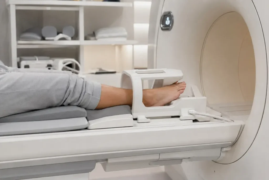
- Positioning. The patient lies on the movable scanner table. The foot and ankle joint are placed in a special coil.
- Leg stabilization. The limb is secured in a comfortable position to prevent movement during scanning.
- Scanning. The scanner acquires a series of cross-sectional images in different planes.
- Contrast enhancement. If contrast-enhanced MRI has been prescribed, the agent is administered intravenously, after which additional image series are acquired.
- Reporting. The radiologist analyzes the images and prepares a report.
The procedure is usually painless. The scanner produces loud rhythmic sounds, so hearing protection is provided. Communication with the radiology technologist is maintained throughout the examination.
It is important to remain still to obtain clear images.
How long an ankle MRI takes
The duration depends on the protocol, the need for contrast enhancement, the number of additional sequences and the patient’s ability to remain still.
An MRI with contrast may take longer because an intravenous catheter must be placed, the contrast agent administered and additional scanning performed. The approximate examination time can be confirmed when booking.
Contraindications and limitations
Before MRI, inform the staff if you have:
- a pacemaker;
- an implanted defibrillator;
- a cochlear implant;
- a neurostimulator;
- an insulin pump;
- a continuous glucose monitoring system;
- vascular clips;
- metal fragments or foreign bodies;
- other electronic or metal implants;
- a recent operation;
- pregnancy;
- severe claustrophobia;
- a condition that prevents you from remaining still.
Most orthopedic implants do not prevent MRI, but they may cause artifacts on the images. The final decision depends on the material, device model and MRI compatibility.
If a plate, screws or other metal hardware has been installed in the ankle area, mention this when booking. The HBMedical price list includes separate MRI options for patients with metal hardware.
What the patient receives after the examination
After scanning, the radiologist analyzes the images and prepares a report.
The patient receives:
- MR images of the ankle joint;
- a radiologist’s report;
- a recording of the examination on a USB flash drive;
- results for consultation with an orthopedic trauma specialist, surgeon or another specialist.
At HBMedical, the report is usually prepared within 5–7 hours. It can be collected in person or accessed through the patient portal. In complex cases, the time may vary.
The MRI report describes the detected changes but does not replace a clinical diagnosis. Further treatment is determined by the relevant specialist.
Why undergo an ankle MRI at HBMedical
- a Siemens Magnetom Sempra 1.5 Tesla scanner;
- examination of the ankle joint and proximal foot;
- MRI without contrast and with contrast enhancement;
- detailed evaluation of the ligaments, tendons, cartilage and bone structures;
- image interpretation by a radiologist;
- the ability to compare the findings with previous examinations;
- a report and recording of the results on a USB flash drive;
- a convenient location near Shuliavska metro station;
- the diagnostic department is open every day;
- advance booking at a convenient time.
Where to get an ankle MRI in Kyiv
You can undergo an ankle MRI at the HBMedical medical center at: 3 Oleksandra Dovzhenka Street, Kyiv, near Shuliavska metro station.
The medical center is located on the right bank of Kyiv. It is convenient for patients from Shuliavka, KPI, Nyvky, Lukianivka, Dorohozhychi and the Shevchenkivskyi, Solomianskyi and Sviatoshynskyi districts.
When booking, inform the administrator of:
- the exact name of the examination specified in the referral;
- which leg needs to be examined;
- the exact location of the pain — in the joint, foot or Achilles tendon area;
- whether contrast enhancement is required;
- whether previous images are available;
- whether any operations have been performed;
- whether metal hardware is present;
- whether you have claustrophobia.
Book an ankle MRI at HBMedical to undergo the examination in Kyiv near Shuliavska and receive a report for further consultation with an orthopedic trauma specialist.
Frequently asked questions
What does an ankle MRI show?
MRI helps evaluate the bones, articular cartilage, ligaments, tendons, synovial membrane and soft tissues. The examination may show ligament tears, tendon injuries, bone marrow edema, occult fractures, osteoarthritis, synovitis and other changes.
How much does an ankle MRI cost in Kyiv?
According to the current HBMedical price list, an MRI of the ankle joint and proximal foot without contrast costs UAH 3,750, while an examination with contrast costs UAH 6,500.
Is the foot included in an ankle MRI?
At HBMedical, the examination includes the ankle joint and the proximal part of the foot. The metatarsal bones and phalanges are covered by a separate distal foot MRI protocol.
Can an ankle ligament tear be seen on MRI?
Yes. MRI helps identify the injured ligament, the location of the injury and the degree of tearing, as well as associated changes in the tendons, cartilage and bones.
Is contrast required?
Contrast is not required for most ligament, tendon and cartilage injuries. It may be prescribed if a tumor, infection, active inflammatory process or complex postoperative changes are suspected.
Can an MRI be performed immediately after an injury?
Yes, if the examination has been prescribed by a doctor. However, X-ray imaging is often the initial method for an acute injury. MRI is performed when the ligaments, tendons, cartilage or an occult bone injury need to be assessed.
How should I prepare for an ankle MRI?
No special preparation is usually required for an examination without contrast. Bring the referral, previous images and implant documentation, if applicable.
Can I have an MRI with a plate or screws in my leg?
This depends on the material and MRI compatibility of the hardware. You must mention the metal hardware when booking and provide the relevant documentation if possible.
How does an ankle MRI differ from an Achilles tendon MRI?
An ankle MRI covers the joint, ligaments, adjacent tendons and bone structures. A separate protocol may be used to evaluate the Achilles tendon in detail throughout the required area.
Are an ankle MRI and a foot MRI the same examination?
Not entirely. The areas partly overlap, but the examinations have different scanning centers. Before booking, provide the exact location of the symptoms and the wording from the referral.
When will the report be ready?
At HBMedical, the report is usually prepared within 5–7 hours. In complex cases, it may take longer.
This material is for informational purposes only and does not replace a medical consultation.










Мені все сподобалось
Всі профі
Дякую


Велика подяка лікарю Дуброву Є.О. — надзвичайно компетентний і чуйний фахівець, який детально все пояснив, розвіяв тривогу та підтримав морально. Щиро дякую анестезіологу Бєлаш О.М. за високий професіоналізм, чуйність і справжнє людське ставлення — занурення в сон та сам перебіг процедури минули максимально комфортно. Також величезне спасибі медсестрі (на жаль, не дізналася ім'я) за її турботу, доброту та увагу.
Весь персонал вразив своєю чуйністю та теплом. Дякую за ваш труд і турботу про пацієнтів! Однозначно рекомендую.














Дуже ввічливий та приємний персонал клініки. Лікар був уважний, без поспіху пройшовся по всім аспектам здоровʼя. Лабораторія швидко зробила аналізи. В цілому дуже задоволена. Радже звертатися!




Саме до таких фахівців, в разі необхідності, є бажання повернутися!

і закінчуючи отриманим описом і рекомендаціями від лікарів.
























Need specialist help?
Order a callback and our consultant will help you choose a doctor and make an appointment at a convenient time for you!