








 Work time
Diagnostics: Mon - Sun, 6:00 - 23:00
Polyclinic: Mon - Sun, 8:00 - 20:00

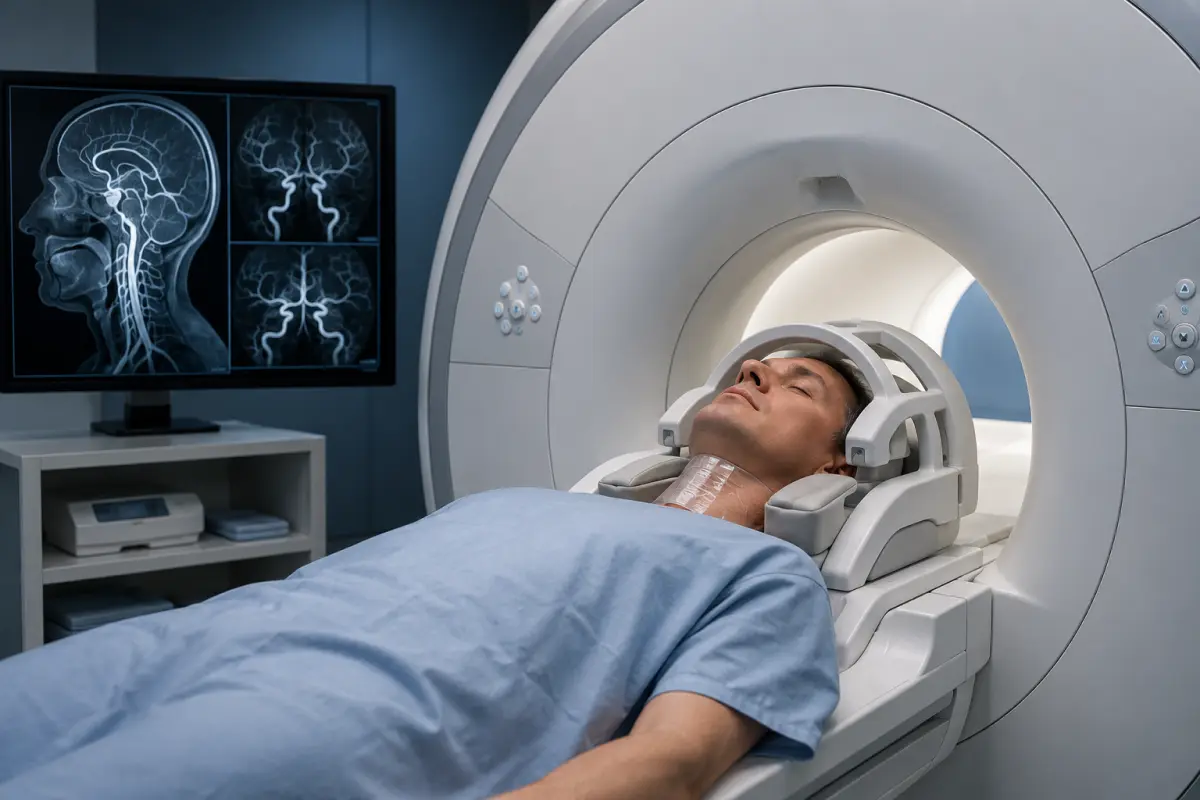
MR angiography is a non-invasive magnetic resonance examination of arteries and veins. The method provides detailed images of blood vessels and helps assess their course, lumen and blood-flow characteristics without using X-ray radiation.
At the HBMedical medical centre, patients can undergo MR angiography of the cerebral vessels, the arteries and veins of the head, as well as the neck vessels. The examination is performed on a Siemens Magnetom Sempra 1.5 Tesla scanner with 3D vascular reconstruction.
Centre address: 3 Oleksandra Dovzhenka Street, Kyiv, near Shuliavska metro station.
According to the current HBMedical price list, the main head and neck MR angiography protocols are performed without intravenous contrast. The exact type of examination should be selected according to the doctor’s referral: MR angiography of the arteries, veins, neck vessels, or a combined MRI of the brain and blood vessels.
Price of MR Angiography in Kyiv
The following examinations are available at HBMedical:
- MR angiography of the cerebral arteries with 3D reconstruction — UAH 3,160;
- MR angiography of the cerebral veins with 3D reconstruction — UAH 3,160;
- MR angiography of the cerebral arteries and veins with 3D reconstruction — UAH 5,660;
- MR angiography of the neck arteries with the aortic arch — UAH 3,160;
- Brain MRI and MR angiography of the arteries — UAH 5,660;
- Brain MRI and MR angiography of the arteries and veins — UAH 7,650;
- Brain MRI, head arteries and neck arteries — UAH 7,650;
- Brain MRI, head arteries and veins, and neck arteries with the aortic arch — UAH 9,550.
The price of an MRI examination of the cerebral vessels depends on which vessels need to be assessed and whether simultaneous imaging of the brain tissue is required. The examination fee includes the radiologist’s report and a copy of the results on a USB drive. The HBMedical price list has been valid since 1 April 2026; however, patients are advised to confirm the current price before their visit.
What Is Angiography?
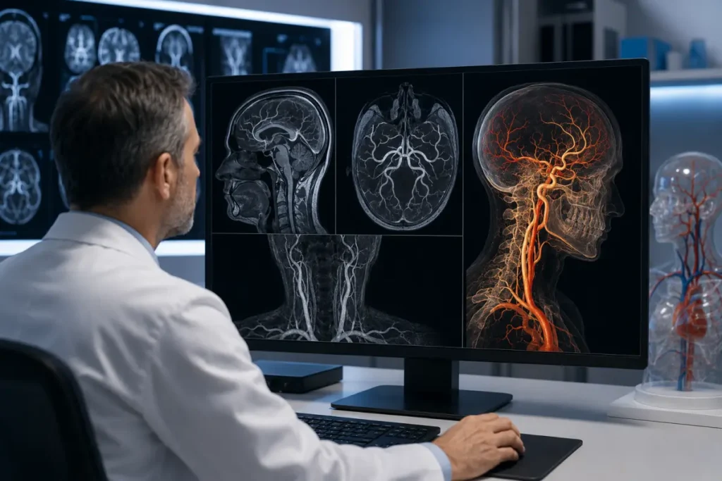
Angiography is a general term for methods used by doctors to examine blood vessels. It can be performed in several ways:
- using magnetic resonance imaging — MR angiography;
- using computed tomography — CT angiography;
- by inserting a catheter directly into a blood vessel — catheter angiography.
This page focuses specifically on magnetic resonance angiography of the cerebral, head and neck vessels. It does not require an arterial puncture and does not use ionising radiation. Depending on the protocol, MR angiography may be performed with or without a contrast agent.
Patients also commonly refer to this examination as:
- MRI of the cerebral vessels;
- MRI of the head vessels;
- MRI of the head and blood vessels;
- MRI angiography of the brain;
- MRI of the head and neck vessels.
What Does an MRI of the Cerebral Vessels Show?
MR angiography provides images of the vascular system and helps assess the structure of arteries or veins within the selected protocol.
The examination may help detect or clarify:
- narrowing of a blood vessel lumen;
- complete blockage or impaired vessel patency;
- aneurysms;
- arteriovenous malformations;
- congenital vascular variations;
- abnormal tortuosity of arteries;
- vascular changes after trauma;
- signs of arterial wall dissection;
- impaired venous drainage;
- thrombosis of veins or venous sinuses;
- changes after surgery or endovascular treatment;
- vascular features important for planning further treatment.
MR angiography helps assess blood vessels but does not always establish the final diagnosis on its own. The findings should be interpreted together with the patient’s symptoms, neurological examination, brain MRI findings and other tests.
MR Angiography of the Cerebral Arteries
MR angiography of the cerebral arteries is intended to assess the intracranial arteries. During the examination, the doctor evaluates their course, diameter, symmetry and patency.
The examination may be used when the following are suspected:
- aneurysm;
- arterial narrowing or occlusion;
- arteriovenous malformation;
- consequences of impaired cerebral circulation;
- congenital vascular abnormality;
- vascular changes after trauma;
- arterial dissection;
- the need to monitor a previously identified condition.
At HBMedical, MR angiography with 3D reconstruction is performed to assess the cerebral arteries. This format helps the doctor analyse blood vessels in different projections.
MR Angiography of the Cerebral Veins
A separate protocol called MR venography is used to examine the veins and venous sinuses. In the price list, it may be referred to as MR angiography of the cerebral veins.
The examination may be recommended when the following are suspected:
- cerebral venous sinus thrombosis;
- impaired venous drainage;
- developmental abnormalities of the veins;
- venous malformations;
- consequences of inflammatory or thrombotic processes;
- changes after surgery or treatment.
MR angiography of the arteries does not replace an examination of the veins. If the doctor needs to assess both systems, the patient should book MR angiography of the cerebral arteries and veins.
MRI of the Head and Neck Vessels
Blood reaches the brain through the neck arteries, so in some clinical situations examining only the intracranial vessels is not sufficient.
MR angiography of the head and neck vessels may include:
- cerebral arteries;
- cerebral veins;
- carotid arteries;
- vertebral arteries;
- neck arteries;
- the aortic arch, depending on the selected protocol.
A comprehensive MRI examination of the head and neck vessels helps assess the vascular pathway from the major neck arteries to the intracranial branches. It may be recommended when stenosis, occlusion, dissection or other changes that can affect the blood supply to the brain are suspected.
When booking, state the exact wording from the referral. Examinations of the «cerebral arteries», «neck arteries» and «head and neck arteries and veins» use different protocols and have different prices.
When Is MR Angiography Prescribed?
A doctor may recommend an MRI of the cerebral vessels when symptoms or conditions may be associated with impaired cerebral circulation.
Possible indications include:
- a previous stroke or transient ischaemic attack;
- temporary weakness or numbness on one side of the body;
- temporary speech disturbances;
- sudden episodes of impaired vision;
- suspected aneurysm;
- a known vascular malformation;
- suspected cerebral venous sinus thrombosis;
- neurological symptoms after a head or neck injury;
- suspected dissection of the carotid or vertebral artery;
- follow-up after surgery or an endovascular procedure;
- the need to clarify changes detected during ultrasound, CT or MRI;
- vascular disease in close relatives, at the doctor’s discretion.
Headache or dizziness alone is not always an indication for MR angiography. The need for the examination is determined by a neurologist or another doctor based on the nature of the symptoms and examination findings.
Brain MRI with Vascular Angiography
Brain MRI and MR angiography are two different but complementary protocols.
Brain MRI shows the brain tissue, ventricular system, meninges and structural changes.
MR angiography is focused on examining the arteries or veins.
A combined brain MRI with vascular angiography may be recommended when the following need to be assessed at the same time:
- brain tissue;
- arteries;
- veins;
- consequences of impaired circulation;
- the relationship between a structural lesion and nearby blood vessels.
If the referral states «MRI of the brain and blood vessels», it is important to clarify which vessels should be examined: the arteries, veins or both systems.
Is Contrast Required for an MRI of the Cerebral Vessels?
MR angiography may be performed with or without a contrast agent. A non-contrast technique based on blood flow within the vessels is often used to visualise the cerebral arteries.
According to the current HBMedical price list, MR angiography of the cerebral arteries, cerebral veins and neck arteries is performed without contrast enhancement. This allows the examination to be carried out without an intravenous injection.
In some clinical situations, the doctor may prescribe a different contrast-enhanced protocol. In that case, the possibility of performing the scan and the preparation requirements should be clarified in advance. Gadolinium-based agents are used for MRI, rather than the iodine-containing contrast usually used for CT angiography.
MR Angiography or CT Angiography: What Is the Difference?
Both methods are used to assess blood vessels, but they work according to different principles.
MR angiography
- uses a magnetic field and radiofrequency pulses;
- does not use ionising radiation;
- may be performed without a contrast agent;
- is suitable for examining the cerebral arteries and veins;
- can be combined with MRI of the brain tissue.
CT angiography
- uses X-ray radiation;
- usually requires an intravenous iodine-containing contrast agent;
- is performed quickly;
- provides detailed images of the vessel lumen;
- is often used in emergency diagnostics.
Catheter angiography is an invasive procedure: a catheter is inserted into a blood vessel, and images are obtained under X-ray guidance. This method may be used not only for diagnosis but also to perform a therapeutic intervention.
The choice between MR angiography, CT angiography and catheter angiography should be made by a doctor.
How to Prepare for MR Angiography
No special preparation is usually required for non-contrast MR angiography of the head and neck vessels. Patients may follow their usual diet and continue taking regular medications unless the doctor has provided other instructions.
Before the examination, it is recommended to:
- bring the doctor’s referral;
- bring previous MRI, CT and vascular ultrasound reports and medical records;
- inform the staff about a pacemaker, defibrillator or other electronic devices;
- bring documents for any implanted devices;
- report any metal fragments or previous operations;
- inform the staff about pregnancy or the possibility of pregnancy;
- mention claustrophobia in advance;
- remove jewellery, piercings, hearing aids and other metal objects;
- not stop prescribed medications without medical advice.
If the doctor has prescribed contrast-enhanced MR angiography, the patient should report kidney disease and any previous reactions to contrast agents. If risk factors are present, a blood creatinine test may be required.
How Is MR Angiography Performed?
Before the procedure, the patient completes a safety questionnaire and leaves metal and electronic items outside the scanning room.
The patient lies on the movable scanner table. The head and neck are positioned in a special coil and may be secured if necessary to reduce movement.
During scanning, the machine creates a series of vascular images in different planes. These images are used to form a 3D reconstruction, which is analysed by the radiologist.
The scanner produces loud rhythmic sounds, so hearing protection is provided. The patient can communicate with the radiographer throughout the procedure.
It is important to remain still to obtain clear images.
How Long Does an MRI of the Cerebral Vessels Take?
The duration depends on the selected protocol and the number of areas examined:
- arteries only;
- veins only;
- arteries and veins;
- head and neck vessels;
- brain MRI combined with angiography.
The approximate duration should be confirmed when booking by giving the administrator the full name of the examination stated in the referral.
Contraindications and Limitations
MR angiography is performed in a strong magnetic field. Before the examination, the staff must be informed if the patient has:
- a pacemaker;
- an implanted cardioverter-defibrillator;
- a cochlear implant;
- a neurostimulator;
- an insulin pump;
- a continuous glucose monitoring system;
- vascular clips;
- metal fragments or foreign bodies;
- middle- or inner-ear implants;
- other electronic or metal medical devices;
- recently undergone surgery;
- pregnancy;
- severe claustrophobia;
- a condition that prevents lying still.
Having an implant does not always mean that MRI is prohibited. Whether scanning is possible depends on the device model and the conditions of its MR compatibility.
What Does the Patient Receive after the Examination?
After scanning, the radiologist analyses the acquired images and 3D vascular reconstructions.
The patient receives:
- MR images of the blood vessels;
- a 3D reconstruction;
- a radiologist’s report;
- a copy of the results on a USB drive;
- materials for consultation with a neurologist, neurosurgeon or vascular specialist.
At HBMedical, the report is usually prepared within 5–7 hours. In complex cases, it may take longer. The report describes the detected changes but does not replace the final clinical diagnosis.
Why Choose MR Angiography at HBMedical?
- Siemens Magnetom Sempra 1.5 Tesla scanner;
- examination of the cerebral arteries and veins;
- MR angiography of the neck vessels;
- combined MRI of the brain and blood vessels;
- 3D reconstruction of the vascular system;
- main protocols performed without contrast;
- image interpretation by a radiologist;
- the possibility of comparison with previous examinations;
- digital results;
- convenient location near Shuliavska metro station;
- appointments available at a convenient time.
Where to Have an MRI of the Cerebral Vessels in Kyiv
MR angiography of the cerebral, head and neck vessels is available at the HBMedical medical centre at 3 Oleksandra Dovzhenka Street, Kyiv, near Shuliavska metro station.
The centre is located on the right bank of Kyiv and is convenient for patients from Shuliavka, KPI, Nyvky, Lukianivka, Dorohozhychi and the Shevchenkivskyi, Solomianskyi and Sviatoshynskyi districts.
When booking, tell the administrator:
- the exact wording from the referral;
- which vessels need to be examined — arteries, veins or neck vessels;
- whether a brain MRI is required;
- whether previous results are available;
- whether any implants or electronic medical devices are present;
- whether the patient has claustrophobia.
Book MR angiography at HBMedical to have the cerebral, head and neck vessels examined in Kyiv near Shuliavska and receive a report for further medical consultation.
Frequently Asked Questions
What is angiography?
Angiography is a group of methods used to examine blood vessels. It may be performed using MRI, CT or X-ray equipment with catheter insertion.
What is MR angiography?
MR angiography is a magnetic resonance examination of arteries and veins. It does not use ionising radiation and, in many cases, can be performed without a contrast agent.
What does an MRI of the cerebral vessels show?
The examination helps assess the course and patency of blood vessels and detect narrowing, occlusions, aneurysms, malformations, cerebral venous sinus thrombosis and other vascular changes.
How much does an MRI of the cerebral vessels cost?
MR angiography of the cerebral arteries at HBMedical costs UAH 3,160. Examination of the arteries and veins costs UAH 5,660. The price of a combined brain and vascular MRI depends on the selected protocol.
Is contrast required for MR angiography?
Not always. According to the current HBMedical price list, the main cerebral and neck MR angiography protocols are performed without contrast.
Are MR angiography and MRI of the cerebral vessels the same?
Yes. In everyday language, these terms are often used interchangeably. The more medically precise term is MR angiography.
How is MR angiography different from brain MRI?
Brain MRI assesses the brain tissue, while MR angiography examines the arteries or veins. If necessary, both protocols can be performed during one comprehensive examination.
Can brain MRI and vascular MRI be performed at the same time?
Yes. HBMedical offers combined brain MRI protocols with MR angiography of the arteries, veins, or head and neck vessels.
How should I prepare for an MRI of the cerebral vessels?
No special preparation is usually required for a non-contrast examination. Bring the referral, previous results and documents for implanted devices.
Is a referral required?
The possibility of undergoing the examination without a referral can be clarified when booking. A referral is advisable because it helps select the correct vessels and scanning protocol.
When will the report be ready?
At HBMedical, the report is usually prepared within 5–7 hours. In complex cases, the timeframe may vary.
This material is for informational purposes only and does not replace medical consultation.










Мені все сподобалось
Всі профі
Дякую


Велика подяка лікарю Дуброву Є.О. — надзвичайно компетентний і чуйний фахівець, який детально все пояснив, розвіяв тривогу та підтримав морально. Щиро дякую анестезіологу Бєлаш О.М. за високий професіоналізм, чуйність і справжнє людське ставлення — занурення в сон та сам перебіг процедури минули максимально комфортно. Також величезне спасибі медсестрі (на жаль, не дізналася ім'я) за її турботу, доброту та увагу.
Весь персонал вразив своєю чуйністю та теплом. Дякую за ваш труд і турботу про пацієнтів! Однозначно рекомендую.














Дуже ввічливий та приємний персонал клініки. Лікар був уважний, без поспіху пройшовся по всім аспектам здоровʼя. Лабораторія швидко зробила аналізи. В цілому дуже задоволена. Радже звертатися!




Саме до таких фахівців, в разі необхідності, є бажання повернутися!

і закінчуючи отриманим описом і рекомендаціями від лікарів.
























Need specialist help?
Order a callback and our consultant will help you choose a doctor and make an appointment at a convenient time for you!