








 Work time
Diagnostics: Mon - Sun, 6:00 - 23:00
Polyclinic: Mon - Sun, 8:00 - 20:00

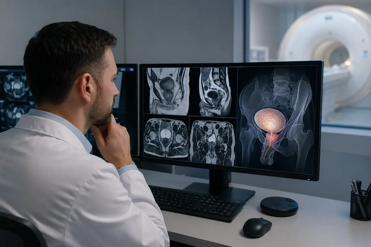
MRI of the male pelvic organs is a detailed magnetic resonance examination of the prostate gland, seminal vesicles, urinary bladder, rectum, lymph nodes and surrounding soft tissues. The examination helps clarify changes detected during ultrasound, laboratory tests or a clinical examination, assess the extent of a pathological process and monitor treatment results.
At the HBMedical medical centre, men can undergo a pelvic MRI in Kyiv without contrast or with intravenous contrast enhancement. The examination is performed on a Siemens Magnetom Sempra 1.5 Tesla scanner at 3 Oleksandra Dovzhenka Street, Kyiv, near Shuliavska metro station.
According to the current HBMedical price list, a male pelvic MRI covers the prostate gland, urinary bladder and rectum. The cost is UAH 5,400 without contrast and UAH 8,400 with contrast enhancement.
You can book an MRI of the male pelvic organs by phone or through the form on the website. When booking, provide the exact wording from the referral and clarify whether contrast enhancement is required.
Price of MRI of the Male Pelvic Organs
- Male pelvic MRI without contrast — UAH 5,400.
- MRI of the male pelvic organs with contrast — UAH 8,400.
The examination price includes MR images, a radiologist’s report and the results recorded on a USB flash drive. Before your visit, we recommend checking the current price because the price list may be updated.
The cost of a separate MRI of the urinary bladder, rectum, scrotum or a combined examination of the abdominal cavity and pelvis may differ. Therefore, when booking, it is important to tell the administrator not only the name of the area but also the clinical question specified by the doctor.
What Is a Male Pelvic MRI?
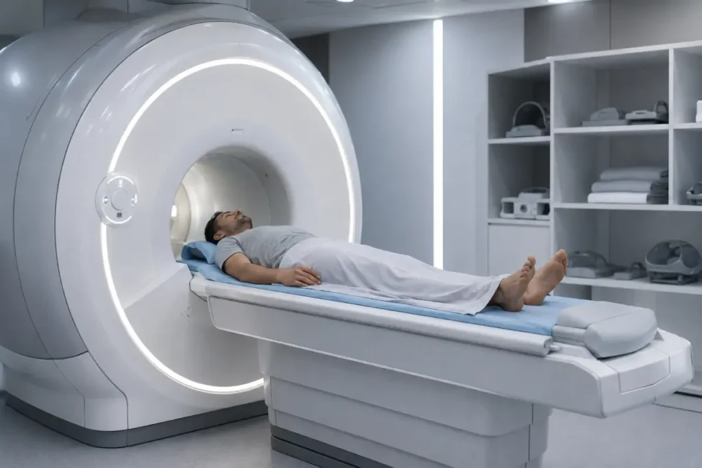
Magnetic resonance imaging uses a strong magnetic field, radiofrequency pulses and computer processing to create cross-sectional images of internal organs. MRI does not use X-rays or any other ionising radiation.
The examination is particularly informative for assessing soft tissues. It helps visualise the structure of organs, the relationship between anatomical structures, lymph nodes and the extent of a pathological process within the pelvis.
MRI does not replace a consultation with a urologist, proctologist, oncologist or another doctor. The radiologist’s report should be assessed together with the symptoms, laboratory test results, ultrasound findings, clinical examination and previous imaging studies.
What Is Included in a Male Pelvic MRI?
During a standard male pelvic MRI at HBMedical, the following are assessed:
- the prostate gland;
- the seminal vesicles;
- the urinary bladder;
- the rectum;
- pelvic lymph nodes;
- soft tissues of the pelvic region;
- surrounding anatomical spaces;
- postoperative and scar-related changes;
- possible spread of an inflammatory or tumour process.
The scope of the examination depends on the referral and selected protocol. If the doctor needs a targeted assessment of the prostate gland, urinary bladder or rectum, a separate specialised protocol may be required.
MRI of the scrotum, testicles and penis is a separate service and is not automatically included in a standard MRI of the male pelvic organs.
What Does MRI of the Male Pelvic Organs Show?
MRI helps detect or clarify structural changes in the organs and tissues of the pelvic region. Depending on the clinical situation, the examination may show:
- enlargement and structural changes of the prostate gland;
- suspicious prostate lesions;
- inflammatory changes in the prostate gland and surrounding tissues;
- changes in the seminal vesicles;
- lesions or thickening of the urinary bladder wall;
- pathological changes in the rectum;
- cysts and other space-occupying lesions;
- inflammatory infiltrates and fluid collections;
- enlarged or altered lymph nodes;
- postoperative and scar-related changes;
- local spread of a tumour process;
- complications after surgery or treatment;
- possible causes of chronic pelvic pain.
A male pelvic MRI cannot always determine the exact nature of a detected lesion on its own. A final diagnosis may require laboratory tests, histological examination, endoscopy, biopsy or consultation with a relevant specialist.
MRI of the Prostate Gland
The prostate gland is one of the main structures assessed during a male pelvic MRI. The doctor may evaluate:
- the size and volume of the prostate;
- the symmetry and contours of the gland;
- the structure of the peripheral and transition zones;
- the presence of suspicious areas;
- the condition of the prostate capsule;
- the seminal vesicles;
- the surrounding fatty tissue;
- pelvic lymph nodes;
- changes after biopsy, surgery, radiotherapy or medical treatment.
A specialised prostate MRI may be used to assess suspicious changes, determine the extent of prostate cancer, look for signs of spread beyond the prostate and plan further diagnosis or treatment.
If the main objective is a detailed examination of the prostate, you should inform the administrator when booking. A standard male pelvic MRI and a specialised prostate protocol may include different imaging sequences.
How Is a Pelvic MRI Different from a Prostate MRI?
MRI of the male pelvic organs covers several organs and structures: the prostate, urinary bladder, rectum, seminal vesicles, lymph nodes and surrounding tissues.
MRI of the prostate gland is a more targeted examination. It may include thinner slices and specialised sequences for a detailed assessment of prostate structure.
When booking, follow the exact wording on the referral:
- «Male pelvic MRI»;
- «MRI of the male pelvic organs»;
- «MRI of the prostate gland»;
- «Prostate MRI with contrast»;
- «Rectal MRI»;
- «Urinary bladder MRI».
It is not recommended to replace one examination with another on your own.
When Is a Male Pelvic MRI Prescribed?
The examination may be prescribed by a urologist, proctologist, oncologist, surgeon, gastroenterologist or family doctor.
Possible indications for MRI of the male pelvic organs include:
- pain or a sensation of pressure in the pelvic area;
- persistent perineal pain;
- urination problems;
- frequent urge to urinate;
- a weak, difficult or intermittent urine stream;
- a feeling of incomplete bladder emptying;
- blood in the urine;
- blood in the semen;
- an elevated PSA level, as determined by a urologist;
- suspected prostate lesion;
- suspected tumour of the urinary bladder or rectum;
- changes detected during ultrasound, CT or digital rectal examination;
- enlarged pelvic lymph nodes;
- preparation for surgery;
- assessment of the extent of a diagnosed disease;
- follow-up after surgical, radiation or medical treatment;
- the need to clarify the cause of chronic pelvic pain.
The presence of one of these symptoms does not necessarily mean that a patient requires MRI. For many urological conditions, the initial diagnostic methods may include a clinical examination, laboratory tests and ultrasound. The doctor determines whether MRI is appropriate.
MRI of the Male Pelvic Organs with Contrast
A pelvic MRI with contrast is performed with intravenous administration of a gadolinium-based agent. Contrast enhancement helps assess tissue blood supply, lesion boundaries and how different areas take up the contrast agent.
Contrast enhancement may be recommended when it is necessary to:
- clarify the nature of a detected lesion;
- assess the prostate gland in greater detail;
- determine the spread of a tumour process;
- assess pelvic lymph nodes;
- examine the wall of the urinary bladder or rectum;
- distinguish a postoperative scar from active changes;
- clarify an inflammatory process;
- monitor the condition after treatment;
- obtain additional information after a non-contrast MRI.
Not every patient requires contrast. The decision depends on the clinical question, referral and previous results.
Before contrast administration, inform the staff about kidney disease and any previous reactions to contrast agents. If risk factors are present, a blood test may be required to assess kidney function. Gadolinium-based agents are used for MRI.
Male Pelvic MRI without Contrast
A non-contrast MRI may be sufficient when it is necessary to:
- assess the general anatomy of the pelvic organs;
- determine the size and structure of the organs;
- detect large space-occupying lesions;
- assess some inflammatory or postoperative changes;
- compare the condition with a previous examination;
- perform an examination using a protocol that does not require contrast enhancement.
Do not decline prescribed contrast enhancement on your own solely because of the price difference. Without contrast, the doctor may not obtain some of the information needed to answer the clinical question.
Preparation for MRI of the Male Pelvic Organs
The exact preparation depends on what needs to be examined: the entire male pelvis, the prostate gland, the urinary bladder or the rectum.
General recommendations:
- bring the doctor’s referral;
- bring the results of previous MRI, CT and ultrasound examinations;
- bring urological test results, PSA levels, medical reports and biopsy results, if available;
- inform the staff about previous surgery and treatment;
- inform the staff about a pacemaker, implants, metal structures and foreign bodies;
- bring documents confirming that your implants are MRI-compatible;
- remove jewellery, watches, bank cards and electronic devices;
- do not stop taking regular medicines without your doctor’s advice;
- clarify the dietary requirements when booking;
- clarify how full the urinary bladder should be;
- inform the staff if you have recently undergone a prostate biopsy, surgery or another procedure.
Instructions regarding food and drink before MRI may vary depending on the protocol and the requirements of the diagnostic centre. Unless you are given other instructions, regular medicines are usually taken as usual.
How to Prepare the Bowel
Bowel movement and pronounced gas formation may cause artefacts on the images. Therefore, before certain protocols, the following may be recommended:
- a light diet the day before;
- temporary restriction of foods that increase gas formation;
- not eating for several hours before the procedure;
- emptying the rectum;
- taking a medication to reduce bowel movement directly at the medical centre.
Do not use laxatives, enemas or antispasmodics on your own. The need for this preparation should be clarified when booking.
How Full Should the Urinary Bladder Be?
Some protocols require a moderately full bladder, while others require an empty bladder. This depends on whether the prostate gland, urinary bladder wall, rectum or the entire pelvis is being assessed.
The administrator should tell you:
- whether you may urinate before the examination;
- how much water you need to drink;
- how long before the procedure you should begin preparation.
Preparation for a Pelvic MRI with Contrast
Before contrast enhancement, you should additionally:
- inform the staff about kidney disease;
- inform the staff about any previous reaction to an MRI contrast agent;
- clarify whether a creatinine blood test is required;
- follow the dietary instructions provided when booking;
- not stop treatment on your own.
How Is a Male Pelvic MRI Performed?
Before the examination, the patient completes a safety questionnaire and leaves metal and electronic items outside the scanning room.
The procedure takes place in several stages:
- Preparation. The patient changes clothes if necessary and lies down on the movable MRI table.
- Positioning. A special receiver coil is placed over the pelvic area, after which the table moves into the scanner.
- Scanning. The scanner creates a series of cross-sectional images in different planes. The patient must remain still and follow the staff’s instructions.
- Contrast enhancement. If an MRI with contrast has been prescribed, the agent is administered through an intravenous catheter, followed by additional scanning sequences.
- Reporting. The radiologist analyses the images and prepares a report.
During scanning, the machine produces loud rhythmic sounds. The patient is provided with hearing protection, and communication with the radiographer is maintained throughout the examination.
How Long Does a Male Pelvic MRI Take?
The duration depends on the selected protocol, the need for contrast enhancement, the number of imaging sequences and the patient’s ability to remain still.
An MRI with contrast may take longer because time is needed to insert the catheter, administer the agent and perform additional scans. The approximate duration should be clarified when booking.
Contraindications and Limitations
A strong magnetic field may affect some medical devices. Be sure to inform the staff if you have:
- a pacemaker;
- an implanted defibrillator;
- a cochlear implant;
- a neurostimulator;
- an insulin pump;
- a continuous glucose monitoring system;
- vascular clips;
- metal fragments or foreign bodies;
- other electronic or metal implants;
- recent surgery;
- severe claustrophobia;
- a condition that prevents you from lying still.
The presence of an implant does not always rule out MRI. The possibility of performing the examination depends on the device model, its MRI compatibility and the conditions specified by the manufacturer.
What Does the Patient Receive after the Examination?
After the scan, the radiologist analyses the images and prepares a report.
The patient receives:
- MR images of the pelvic organs;
- a radiologist’s report;
- the examination recorded on a USB flash drive;
- materials for consultation with a urologist, proctologist, oncologist or another specialist.
The MRI report describes the detected changes but does not replace a clinical diagnosis. Further steps are determined by the doctor who referred the patient for the examination.
Pelvic MRI, Ultrasound or CT
Ultrasound
Ultrasound is often used as the initial method for assessing the prostate gland, urinary bladder and urinary system. It is quick and accessible, but it does not always provide sufficient information about deep soft tissues and the extent of a pathological process.
MRI
MRI provides detailed images of soft tissues and helps assess the prostate, seminal vesicles, urinary bladder, rectum, lymph nodes and surrounding structures.
CT
CT is faster and may be useful for assessing bones, stones, trauma, acute conditions or for a combined examination of the abdominal cavity and pelvis. The method uses ionising radiation.
The choice between MRI, CT and ultrasound should be made by a doctor depending on the clinical question.
Why Have an MRI at HBMedical?
- examination on a Siemens Magnetom Sempra 1.5 Tesla scanner;
- male pelvic MRI without contrast and with contrast enhancement;
- assessment of the prostate gland, urinary bladder and rectum;
- image interpretation by a radiologist;
- the possibility of comparison with previous examinations;
- results recorded on a USB flash drive;
- convenient location near Shuliavska metro station;
- advance booking online or by phone.
Where to Have an MRI of the Male Pelvic Organs in Kyiv
Men can undergo a pelvic MRI at the HBMedical medical centre at 3 Oleksandra Dovzhenka Street, Kyiv, near Shuliavska metro station.
The location is convenient for patients from Shuliavka, KPI, Lukianivka, Dorohozhychi, Nyvky, and the Shevchenkivskyi, Solomianskyi and Sviatoshynskyi districts of Kyiv.
When booking, tell the administrator:
- the exact name of the examination from the referral;
- whether contrast enhancement is required;
- whether a specialised prostate protocol is needed;
- whether you have previous MRI, CT, ultrasound or biopsy results;
- whether you have any implants;
- whether you have kidney disease;
- whether you have claustrophobia.
Book an MRI of the male pelvic organs at HBMedical to undergo the examination in Kyiv near Shuliavska and receive the results for a follow-up consultation with your doctor.
Frequently Asked Questions
What Is Included in a Male Pelvic MRI?
During a male pelvic MRI at HBMedical, the prostate gland, urinary bladder and rectum are assessed. Depending on the protocol, the seminal vesicles, lymph nodes, surrounding soft tissues and postoperative changes are also evaluated.
What Does MRI of the Male Pelvic Organs Show?
The examination may show structural changes in the prostate, urinary bladder and rectum, space-occupying lesions, inflammatory processes, altered lymph nodes and the extent of a pathological process.
How Much Does a Male Pelvic MRI Cost?
According to the current HBMedical price list, an MRI without contrast costs UAH 5,400, while an MRI with contrast costs UAH 8,400. We recommend checking the current price before booking.
When Is a Pelvic MRI with Contrast Required?
Contrast may be required to clarify the nature of a lesion, assess the prostate and lymph nodes, determine tumour spread, or evaluate inflammation and postoperative changes.
How Should a Man Prepare for a Pelvic MRI?
Bring the referral and previous results, clarify the dietary instructions and required bladder filling, inform the staff about implants, and do not take laxatives or antispasmodics on your own.
Can I Eat before the MRI?
This depends on the protocol. In some cases, you may eat as usual; in others, you may be asked not to eat for several hours. You should obtain precise instructions when booking.
Is Bowel Cleansing Required?
Not for every examination. Bowel emptying may be recommended before certain prostate or rectal MRI protocols. Do not use an enema or laxatives without instructions from the medical centre.
Is a Creatinine Test Required?
A creatinine test may be required before an MRI with contrast, especially if you have kidney disease or other risk factors.
How Is a Male Pelvic MRI Performed?
The patient lies on the MRI table while the scanner obtains a series of cross-sectional images of the pelvic region. The patient must remain still during the procedure. If necessary, a contrast agent is administered intravenously.
Are the Testicles Included in a Pelvic MRI?
No. MRI of the scrotum, testicles and penis is a separate examination and requires a dedicated protocol.
Are a Pelvic MRI and a Prostate MRI the Same?
Not exactly. A pelvic MRI covers several organs, while a prostate MRI is a targeted examination of the prostate gland. The required protocol is determined by the doctor’s referral.
Can I Have an MRI without a Referral?
You can clarify whether the examination is available without a referral when booking. A referral is recommended because it helps select the correct area and scanning protocol.
This material is for informational purposes only and does not replace a consultation with a doctor.










Мені все сподобалось
Всі профі
Дякую


Велика подяка лікарю Дуброву Є.О. — надзвичайно компетентний і чуйний фахівець, який детально все пояснив, розвіяв тривогу та підтримав морально. Щиро дякую анестезіологу Бєлаш О.М. за високий професіоналізм, чуйність і справжнє людське ставлення — занурення в сон та сам перебіг процедури минули максимально комфортно. Також величезне спасибі медсестрі (на жаль, не дізналася ім'я) за її турботу, доброту та увагу.
Весь персонал вразив своєю чуйністю та теплом. Дякую за ваш труд і турботу про пацієнтів! Однозначно рекомендую.














Дуже ввічливий та приємний персонал клініки. Лікар був уважний, без поспіху пройшовся по всім аспектам здоровʼя. Лабораторія швидко зробила аналізи. В цілому дуже задоволена. Радже звертатися!




Саме до таких фахівців, в разі необхідності, є бажання повернутися!

і закінчуючи отриманим описом і рекомендаціями від лікарів.
























Need specialist help?
Order a callback and our consultant will help you choose a doctor and make an appointment at a convenient time for you!