








 Work time
Diagnostics: Mon - Sun, 6:00 - 23:00
Polyclinic: Mon - Sun, 8:00 - 20:00

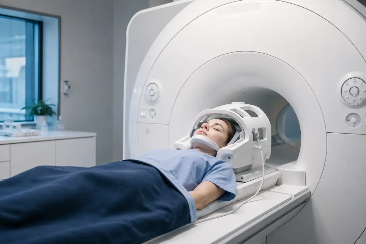
Orbital MRI is a magnetic resonance imaging examination of the eye sockets. It shows the eyeballs, optic nerves, extraocular muscles, blood vessels, fat tissue and lacrimal glands. It is prescribed when it is necessary to find the cause of reduced vision, double vision, pain behind the eye or a bulging eye, and to assess a mass in the orbital area.
At HBMedical, an orbital and optic nerve MRI costs 3200 UAH without contrast and 6000 UAH with contrast. The examination is performed on a Siemens Magnetom Sempra 1.5 Tesla scanner at: Kyiv, 3 Oleksandra Dovzhenka St., 2 minutes from «Shuliavska» metro station. Diagnostics are available every day from 6:00 to 23:00.
Orbital MRI Price
The price depends on three things: whether contrast is needed, whether the brain needs to be examined together with the orbits, and whether the examination is performed under sedation.
If you already have an MRI done at another center, HBMedical radiologists can prepare a report based on those images (2000 UAH). The report can also be issued in English (2000 UAH).
An autumn promotion on MRI is running, depending on the time of the examination: −25% from 5:30 to 7:00, −15% from 7:00 to 8:00 and −20% from 21:00 to 23:30. Please confirm the terms with the call center.
What an Eye and Orbital MRI Shows
MRI distinguishes soft tissues well, so the doctor can see all the structures of the orbit separately. The examination helps to assess:
- the optic nerve along its entire length: thickness, inflammation, compression, masses in it or in its sheaths;
- the optic chiasm, where the fibers of the optic nerves of both eyes cross;
- the extraocular muscles: thickening, swelling, inflammatory changes;
- tumors of the orbit and eyeball, their size and borders;
- inflammatory processes in the orbital fat;
- vascular lesions and dilation of the orbital veins;
- the lacrimal glands;
- the consequences of soft tissue injuries and postoperative changes.
MRI has its limits. The method does not replace an examination by an ophthalmologist: the condition of the retina, the fundus and the transparent media of the eye is assessed with specialized ophthalmological methods. Fractures of the bony walls of the orbit, small calcifications and foreign bodies are shown more reliably by CT.
When an Orbital MRI Is Prescribed
A referral is usually issued by an ophthalmologist, neurologist, neurosurgeon, endocrinologist or ENT doctor. Typical reasons:
- sudden or gradual loss of vision, especially on one side;
- pain behind the eye that gets worse with eye movements;
- double vision, limited eye movement;
- a bulging eyeball (exophthalmos);
- swelling of the optic disc found during a fundus examination;
- suspected optic neuritis, including during an examination for multiple sclerosis;
- thyroid eye disease (endocrine ophthalmopathy) in thyroid disorders;
- a mass noticed on eye ultrasound, CT or during an examination;
- suspected inflammation of the orbital tissues;
- follow-up after treatment or surgery.
Having a symptom does not yet mean that an MRI is what you need. The examination method and its scope are determined by the doctor.
Orbital MRI with or without Contrast
In an MRI with contrast, a gadolinium-based agent is injected into a vein. It accumulates in tissues with active blood flow or inflammation. This helps to see active inflammation of the optic nerve, clarify the borders and nature of a mass and distinguish scar changes from an active process.
Without contrast, the anatomy of the orbit, the size of the muscles, the thickness of the nerve and large space-occupying changes are clearly visible. For some clinical questions, this is sufficient.
Follow the wording in the referral. If the doctor specified «with contrast», do not replace the examination with a non-contrast one because of the price difference: some of the necessary information may be lost.
Before contrast is given, tell staff about kidney disease and any previous reactions to contrast agents. If there are risk factors, a blood test for creatinine may be needed.
Orbits Alone or Together with the Brain
Vision depends on more than the eye. The visual signal travels along the nerve through the chiasm to the visual centers of the brain, so a problem can occur at any point along this pathway. That is why, for some symptoms, such as suspected multiple sclerosis or loss of visual fields, the doctor prescribes a combined examination of the brain, orbits and optic nerves.
If your referral specifies only the brain, see the page brain MRI in Shuliavka. If the doctor suspects changes in the area of the sella turcica, which borders the chiasm, a pituitary MRI with thin slices may be needed.
MRI or CT of the Orbits
| Method | What it shows best | Limitations |
| Orbital MRI | The optic nerve, muscles, fat tissue, soft tissue masses, inflammation | Shows bones and calcifications less well; not performed with metal particles in the eye |
| Orbital CT | Fractures of the orbital walls, calcifications, metallic and other foreign bodies | Uses X-ray radiation, shows soft tissues in less detail |
After a facial injury or when a foreign object in the eye is suspected, the work-up usually starts with CT. MRI is more often chosen to assess the optic nerve and soft tissues.
Safety: Metal in the Eye and on the Eyelids
Before an orbital MRI, it is especially important to tell staff about any metal particles in the eye area. Under the influence of the magnetic field, a metal fragment can move and damage the eye.
Tell the administrator and the MRI technologist if:
- you have worked with welding, grinding or metalworking, even if it was a long time ago;
- you have had an eye injury from a metal object;
- you have shrapnel wounds to the head or face.
If necessary, the doctor may recommend an orbital X-ray first. At HBMedical, it is available on site.
Also tell staff about a pacemaker, cochlear implant, neurostimulator, vascular clips and other implanted devices. Bring the documents confirming their MRI compatibility.
Preparing for an Orbital MRI
A special diet is usually not needed for an MRI without contrast. Before the examination:
- bring your referral and previous results: an ophthalmologist’s report, eye ultrasound, CT or MRI;
- do not apply mascara, eye shadow or eyeliner to your eyelids: cosmetics may contain metal particles that distort the image and may irritate the skin;
- mention permanent eyelid makeup, if you have it;
- remove contact lenses, glasses, jewelry and removable dentures with metal;
- if contrast is prescribed, confirm when booking whether you need to come on an empty stomach and whether a creatinine test is needed;
- do not stop taking regular medications without consulting your doctor.
How the Examination Is Performed
- You fill in a safety questionnaire and leave metal and electronic items outside the room.
- You lie on your back, and your head is placed in a special coil and secured with soft pads.
- The table moves into the scanner. During scanning, you are usually asked to keep your eyes closed and not to move them, because eyeball movements blur the image.
- If contrast is prescribed, the agent is injected into a vein and additional series are acquired.
The procedure is painless, but the machine is loud, so patients are provided with hearing protection. An MRI usually takes 20–40 minutes, and longer with contrast. The scanner is designed for patients weighing up to 220 kg.
At HBMedical, MRI is performed for both adults and children. For young children and people with pronounced claustrophobia who find it hard to lie still, the examination can be performed under sedation.
Results
A radiologist analyzes the images and prepares a report. You can receive it in 5–7 hours, either in person or through your patient account on the website. The images are saved to a USB flash drive so that you can show them to your doctor.
The MRI report describes the detected changes, but the diagnosis is made by the doctor who referred you for the examination. The doctor compares the MRI results with your symptoms and with the findings of the fundus examination, visual field testing and lab tests.
Orbital MRI near «Shuliavska» Metro Station
- Siemens Magnetom Sempra 1.5 Tesla scanner, closed-bore type, manufactured in 2021.
- Diagnostics are available every day from 6:00 to 23:00, so you can come for the examination early in the morning or in the evening.
- Address: 3 Oleksandra Dovzhenka St., a 2-minute walk from «Shuliavska» station. Convenient for residents of Shuliavka, KPI, Lukianivka, Dorohozhychi, Nyvky, and the Shevchenkivskyi, Solomianskyi and Sviatoshynskyi districts.
- There is parking near the clinic.
- For people with special needs, lifts, wide corridors and accessible restrooms are provided. Medical transfer across Kyiv is available.
- The polyclinic, laboratory, X-ray, CT and ultrasound departments work at one center. If the doctor prescribes additional examinations, you will not need to travel to another facility.
- The center has its own independent power supply and operates regardless of whether there is electricity in the city.
For other examination areas and the general price list, see the page MRI in Kyiv at HBMedical.
How to Book
Call +38 (099) 344-30-00 or submit a request on the website, and a consultant will call you back to confirm.
When booking, tell staff:
- the exact name of the examination from the referral: orbits only or orbits with the brain, with or without contrast;
- whether you have had eye injuries or worked with metal;
- whether you have implants, kidney disease or claustrophobia.
Frequently Asked Questions
How much does an orbital MRI cost at HBMedical?
An orbital and optic nerve MRI costs 3200 UAH without contrast and 6000 UAH with contrast. A combined MRI of the brain, orbits and optic nerves costs 5900 UAH without contrast and 7900 UAH with contrast. The report and USB flash drive are included in the price.
What does an MRI of the eye orbits show?
The condition of the optic nerves and chiasm, extraocular muscles, fat tissue, lacrimal glands, blood vessels and eyeballs. The examination also shows masses and inflammatory changes in the orbit.
Are an eye MRI and an orbital MRI the same thing?
Yes. «Eye MRI» is the everyday name for the examination that is called an orbital MRI in medical documentation. It covers both the eyeball and the tissues around it.
Is preparation needed?
For an MRI without contrast, there is usually no special preparation. Come without makeup on your eyelids and remove contact lenses. Before contrast, confirm the eating rules and whether a creatinine test is needed.
Can I have an eye MRI while wearing contact lenses?
It is better to remove lenses before the procedure. During the examination, the eyes are kept closed, and lenses may cause discomfort.
Why do I need to keep my eyes closed during an MRI?
Eyeball movements blur the image. Closed eyes help to keep the gaze still.
Can I have an MRI if a metal shaving once got into my eye?
You must tell staff about this before the examination. A metal fragment can move in a magnetic field. The doctor will determine whether a preliminary X-ray or orbital CT is needed.
How long does an orbital MRI take?
Usually 20–40 minutes. With contrast, the examination takes longer.
When will the result be ready?
The report is prepared in 5–7 hours. You can receive it in person or through your patient account on the website.
Is an orbital MRI performed for children?
Yes, at HBMedical MRI is performed for children as well. If it is hard for a child to lie still, the examination can be performed under sedation.
Does MRI replace an ophthalmologist’s examination?
No. MRI shows the structures of the orbit and the optic nerve, while the condition of the retina and fundus is assessed by an ophthalmologist during an examination.
The information on this page is for reference only and does not replace a doctor’s consultation.





















Мені все сподобалось
Всі профі
Дякую


Велика подяка лікарю Дуброву Є.О. — надзвичайно компетентний і чуйний фахівець, який детально все пояснив, розвіяв тривогу та підтримав морально. Щиро дякую анестезіологу Бєлаш О.М. за високий професіоналізм, чуйність і справжнє людське ставлення — занурення в сон та сам перебіг процедури минули максимально комфортно. Також величезне спасибі медсестрі (на жаль, не дізналася ім'я) за її турботу, доброту та увагу.
Весь персонал вразив своєю чуйністю та теплом. Дякую за ваш труд і турботу про пацієнтів! Однозначно рекомендую.













Дуже ввічливий та приємний персонал клініки. Лікар був уважний, без поспіху пройшовся по всім аспектам здоровʼя. Лабораторія швидко зробила аналізи. В цілому дуже задоволена. Радже звертатися!




Саме до таких фахівців, в разі необхідності, є бажання повернутися!

і закінчуючи отриманим описом і рекомендаціями від лікарів.























Need specialist help?
Order a callback and our consultant will help you choose a doctor and make an appointment at a convenient time for you!