








 Work time
Diagnostics: Mon - Sun, 6:00 - 23:00
Polyclinic: Mon - Sun, 8:00 - 20:00

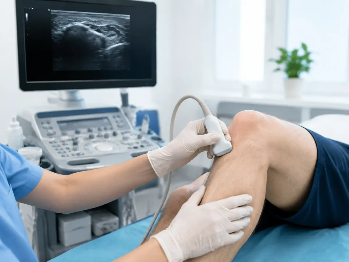
Knee ultrasound is a non-invasive ultrasound examination that allows the doctor to assess the soft tissues of the knee, tendons, ligaments, joint capsule, synovial membrane, and the presence of fluid in the joint.
Knee ultrasound can be useful for pain, swelling, injury, limited mobility, or suspected synovitis, bursitis, or a Baker’s cyst. The method allows the doctor to examine tissues in real time and, if needed, assess them during movement.
You can have the examination done at the HBMedical medical center in Kyiv, in Shuliavka.
Price of knee ultrasound — 650 UAH.
Address: Kyiv, 3 Oleksandra Dovzhenka Street
Metro: “Shuliavska”
Other ultrasound examinations are available on the ultrasound in Kyiv page.
What a Knee Ultrasound Shows
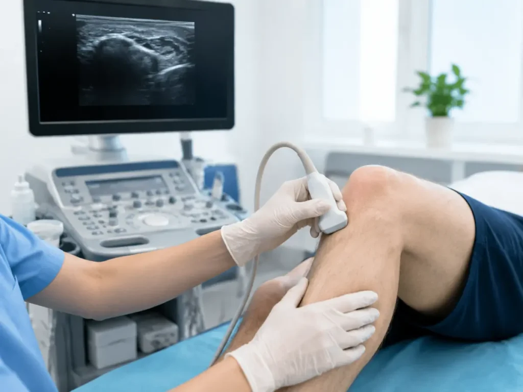
Ultrasound diagnostics is particularly well suited to assessing superficially located soft tissues and structures of the knee.
During the examination, the doctor can assess:
- tendons and their attachment points;
- the collateral ligaments of the knee joint;
- the joint capsule;
- the synovial membrane;
- the amount of fluid in the joint;
- the joint bursae;
- the areas of cartilage accessible to ultrasound;
- the peripheral parts of the menisci;
- the tissues of the popliteal area;
- a Baker’s cyst;
- signs of synovitis;
- signs of bursitis;
- traumatic changes in the soft tissues.
The examination can help detect a joint effusion — an excess buildup of fluid in the knee — inflammatory changes, and damage to certain tendons and superficial ligaments.
Ultrasound is also used to monitor certain changes over time and to track treatment results.
At the same time, ultrasound has its limitations. For a detailed assessment of deep, internal joint structures, including the menisci and cruciate ligaments, the doctor may recommend an MRI.
When You Need a Knee Ultrasound
An ultrasound examination may be needed for either acute symptoms after an injury or gradually developing changes.
It’s worth seeing a doctor if you have:
- knee pain during movement or at rest;
- swelling or puffiness;
- a feeling of stiffness;
- limited bending or straightening;
- clicking in the knee together with pain or other symptoms;
- discomfort after sports or physical exertion;
- a knee injury;
- pain in the area of the ligaments or tendons;
- swelling in the popliteal area;
- suspected synovitis or bursitis;
- the need to monitor previously detected changes.
Clicking without pain or other symptoms doesn’t necessarily indicate a condition, so it’s best to decide whether an examination is needed together with a doctor.
Knee Ultrasound After an Injury
After a blow, a fall, a sports injury, or a sudden movement, a knee ultrasound can help assess the tendons, superficial ligaments, soft tissues, and the presence of fluid in the joint or a hematoma.
But ultrasound isn’t suited to every injury.
If, after the injury:
- you can’t put weight on the leg normally;
- the knee is significantly deformed;
- marked swelling has appeared;
- the joint locks;
- there’s a feeling of instability;
- the pain is very severe,
it’s best to first see an orthopedic traumatologist. The doctor will determine what’s more informative for your specific situation — ultrasound, X-ray, or MRI.
At HBMedical, you can book an orthopedic traumatologist consultation.
Conditions and Changes a Knee Ultrasound Can Show
Depending on the clinical situation, an ultrasound examination can help detect signs of:
- Synovitis. This is inflammation of the synovial membrane, which can be accompanied by fluid buildup in the joint.
- Bursitis. When a joint bursa is inflamed, an ultrasound can assess its size, contents, and surrounding tissues.
- A Baker’s cyst. This is a fluid-filled formation in the popliteal area that is well accessible to ultrasound imaging.
- Tendon and ligament damage. Ultrasound helps assess accessible tendons and superficial ligament structures of the knee.
- Inflammatory and degenerative changes. Ultrasound diagnostics can be used as part of a work-up for arthritis, osteoarthritis, and other joint conditions.
You shouldn’t diagnose yourself based on an ultrasound description. The report needs to be assessed together with your symptoms, examination, and results of other tests.
How a Knee Ultrasound Is Performed
No special preparation is needed for the procedure.
The patient uncovers the knee area and lies down on the examination table. The doctor applies ultrasound gel to the skin and systematically examines the joint with the probe.
The knee is examined from several angles:
- from the front;
- from the inner side;
- from the outer side;
- in the popliteal area.
If needed, the doctor may ask you to bend or straighten your knee to assess certain structures during movement. This ability to examine the joint dynamically is one of the advantages of ultrasound diagnostics.
After the examination is complete, the patient receives the ultrasound doctor’s report.
Do You Need to Prepare for a Knee Ultrasound
No preparation is needed. You can eat, drink water, and follow your usual routine before the examination.
It’s a good idea to:
- wear clothing that makes it easy to uncover the knee joint;
- bring your doctor’s referral, if you have one;
- bring previous ultrasound results;
- bring MRI or X-ray images and reports, if you’ve had them;
- tell the doctor exactly where your knee hurts and which movements cause discomfort.
If the examination is a follow-up, previous results will help assess changes over time.
Ultrasound or MRI of the Knee — What’s the Difference
Ultrasound and MRI address different diagnostic tasks.
Ultrasound is well suited to assessing:
- tendons;
- superficial ligaments;
- joint bursae;
- the synovial membrane;
- fluid in the joint;
- a Baker’s cyst;
- certain soft tissue changes.
Knee MRI shows deep, internal joint structures in more detail, including the menisci, the anterior and posterior cruciate ligaments, the articular cartilage, and the bone marrow.
That’s why ultrasound alone may not be sufficient if a meniscus or cruciate ligament tear is suspected.
If your doctor has recommended a more detailed examination of the internal knee structures, you can have a knee MRI at HBMedical.
Ultrasound or X-ray of the Knee
X-ray and ultrasound also don’t replace each other.
X-ray is primarily used to assess bone structures — for example, when a fracture or degenerative changes are suspected.
Knee ultrasound is more focused on soft tissues — tendons, superficial ligaments, the synovial membrane, joint bursae, and fluid.
Which examination makes sense depends on your symptoms and the results of the physical exam.
Price of Knee Ultrasound in Kyiv
The price of a knee ultrasound at HBMedical is 650 UAH.
| Service | Price |
| Knee ultrasound | 650 UAH |
The current price is listed in HBMedical’s price list.
You can view prices for other examinations on the HBMedical prices page.
If you need both knees examined, let us know when booking and confirm the cost of the examination.
What to Do After the Ultrasound
If the examination was done because of pain, injury, swelling, or limited movement, it’s a good idea to show the report to an orthopedic traumatologist.
The doctor will compare the ultrasound results with your symptoms and examination and determine whether treatment, rehabilitation, or further testing is needed.
HBMedical has an orthopedics and traumatology department.
Where to Get a Knee Ultrasound in Kyiv
You can get a knee ultrasound examination in Kyiv at the HBMedical medical center in Shuliavka.
Address: Kyiv, 3 Oleksandra Dovzhenka Street
Metro: “Shuliavska”
Price: 650 UAH
HBMedical combines ultrasound diagnostics, MRI, and an orthopedics and traumatology department in one medical center.
Frequently Asked Questions
Can a torn meniscus be seen on ultrasound?
Ultrasound only allows assessment of the accessible peripheral parts of the meniscus. For a full assessment of the menisci and to confirm a suspected tear, the doctor more often recommends an MRI.
Can a Baker’s cyst be seen on ultrasound?
Yes. The popliteal area is well accessible to ultrasound, so ultrasound is often used to detect and assess a Baker’s cyst.
Can an ultrasound be done for arthritis?
Yes. Ultrasound can be used to assess the synovial membrane, effusion, and other changes in inflammatory joint conditions. The result needs to be interpreted together with clinical data.
Can a knee ultrasound be done with a knee replacement or metal implants?
Metal implants don’t limit ultrasound the way they do some other imaging methods. However, they can locally affect the accessibility of certain tissues for examination, so be sure to tell the doctor about any surgeries or implants.
Can an ultrasound be done right after an injury?
Yes, if the doctor considers an ultrasound appropriate. It can help assess the soft tissues and the presence of fluid. If a fracture or a deep internal joint injury is suspected, an X-ray or MRI may be needed.
Book a Knee Ultrasound at HBMedical
Need a knee ultrasound in Kyiv? Book an examination at HBMedical near “Shuliavska” metro station.
Knee ultrasound — 650 UAH
Kyiv, 3 Oleksandra Dovzhenka Street
No special preparation is needed. If you have previous knee ultrasound, X-ray, or MRI results, bring them with you.
You can book by phone or through the HBMedical online form.





Мені все сподобалось
Всі профі
Дякую


Велика подяка лікарю Дуброву Є.О. — надзвичайно компетентний і чуйний фахівець, який детально все пояснив, розвіяв тривогу та підтримав морально. Щиро дякую анестезіологу Бєлаш О.М. за високий професіоналізм, чуйність і справжнє людське ставлення — занурення в сон та сам перебіг процедури минули максимально комфортно. Також величезне спасибі медсестрі (на жаль, не дізналася ім'я) за її турботу, доброту та увагу.
Весь персонал вразив своєю чуйністю та теплом. Дякую за ваш труд і турботу про пацієнтів! Однозначно рекомендую.













Дуже ввічливий та приємний персонал клініки. Лікар був уважний, без поспіху пройшовся по всім аспектам здоровʼя. Лабораторія швидко зробила аналізи. В цілому дуже задоволена. Радже звертатися!




Саме до таких фахівців, в разі необхідності, є бажання повернутися!

і закінчуючи отриманим описом і рекомендаціями від лікарів.
























Need specialist help?
Order a callback and our consultant will help you choose a doctor and make an appointment at a convenient time for you!