








 Work time
Diagnostics: Mon - Sun, 6:00 - 23:00
Polyclinic: Mon - Sun, 8:00 - 20:00

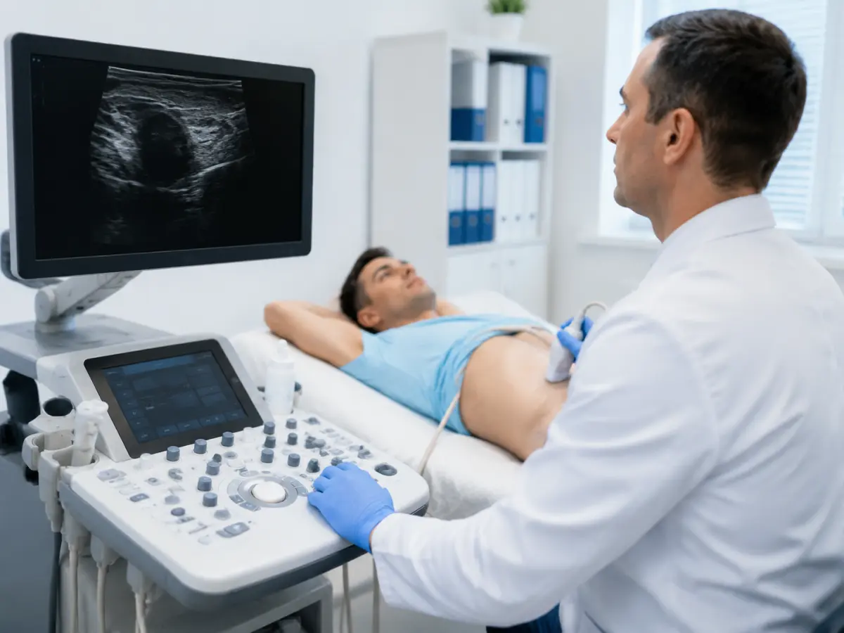
Scrotal ultrasound is an ultrasound examination of the testicles, their epididymides, membranes, and other structures in this area. The examination helps assess the size and structure of the testicles and detect fluid buildup, inflammatory, vascular, traumatic, and other changes. If needed, Doppler mode is used to assess blood flow.
You can get a scrotal ultrasound in Kyiv at the HBMedical medical center in Shuliavka.
Scrotal ultrasound — 850 UAH
Address: Kyiv, 3 Oleksandra Dovzhenka Street
Metro: “Shuliavska”
The current price for the service in HBMedical’s price list is 850 UAH.
Other types of ultrasound diagnostics can be viewed on the ultrasound in Kyiv page.
What Scrotal Ultrasound Is and What the Doctor Examines
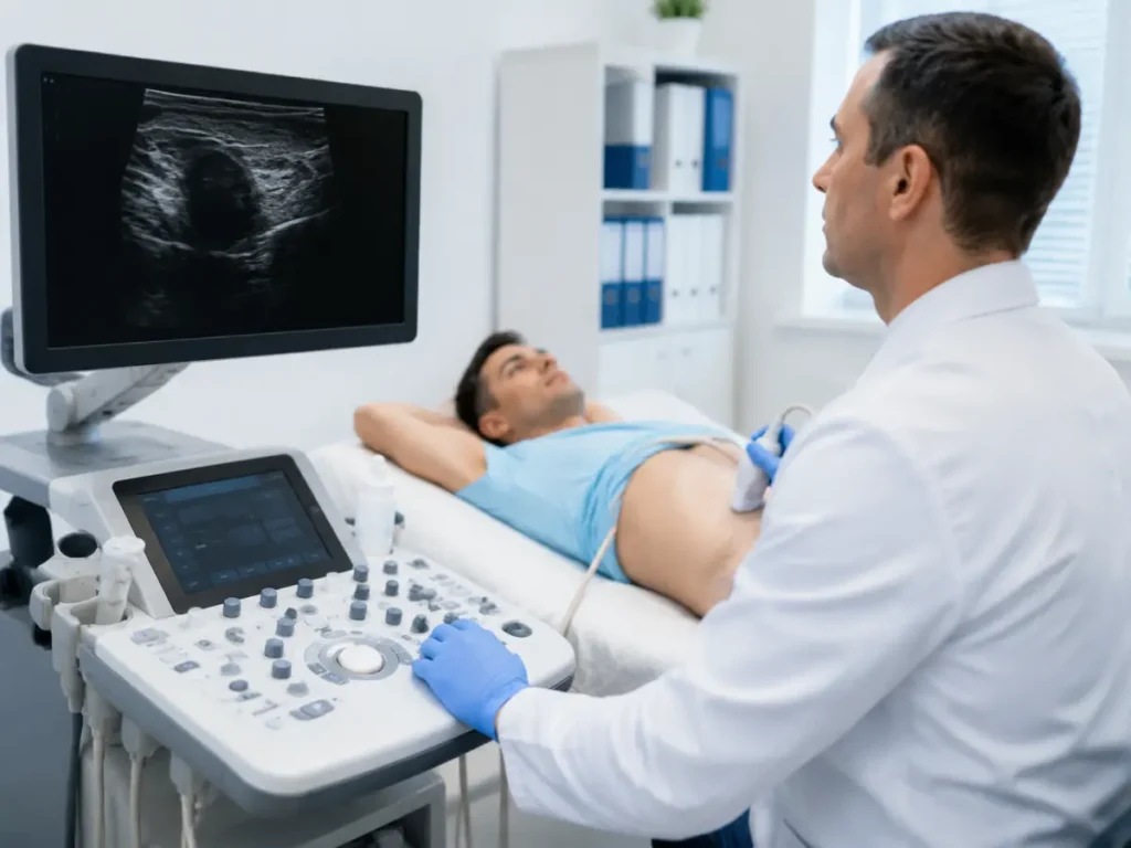
During a testicular ultrasound, the doctor obtains an image of the internal structures using high-frequency sound waves. No ionizing radiation is used. The probe is moved over the skin after ultrasound gel has been applied.
During the examination, the following may be assessed:
- the right and left testicle;
- the size, shape, and structure of the testicles;
- the epididymides;
- the testicular membranes;
- the presence of fluid;
- the spermatic cords and accessible superficial structures;
- any formations or lumps;
- the effects of an injury;
- if indicated — blood flow in the vessels.
The doctor usually compares the right and left sides, even if symptoms occur on only one side.
What Scrotal Ultrasound Shows
Ultrasound helps detect or clarify various structural changes in the testicles and epididymides.
The examination may show signs of:
Varicocele
Varicocele is the dilation of the veins of the spermatic cord. Doppler ultrasound can be used to assess venous blood flow, and in some cases, an examination during straining.
Hydrocele
Hydrocele, or a fluid-filled sac around the testicle, involves a buildup of fluid around the testicle. Ultrasound allows the doctor to see the fluid and assess other structures of the scrotum.
Orchitis and Epididymitis
When the testicle or its epididymis is inflamed, ultrasound can show structural changes, tissue enlargement, and changes in blood flow. Ultrasound is used as part of a comprehensive work-up when orchitis or epididymitis is suspected.
Epididymal Cysts
Ultrasound is well suited to assessing fluid-filled formations and helps determine the location and size of an epididymal cyst.
Effects of an Injury
After a blow or other injury, ultrasound can help assess the testicle, surrounding tissue, hemorrhage, fluid buildup, and other changes accessible to ultrasound.
Testicular Mass
If the patient or doctor has found a lump or nodule in the testicle, an ultrasound examination helps determine its location and characteristics.
When a testicular tumor is suspected, ultrasound is an important initial imaging method. The European Association of Urology recommends high-frequency testicular ultrasound for the diagnostic assessment of suspicious changes.
At the same time, a single ultrasound report doesn’t establish a final histological diagnosis. If a suspicious mass is found, a urologist’s consultation and, if indicated, further tests are needed.
When You Should Get a Testicular Ultrasound
A doctor may recommend the examination when symptoms appear or to clarify changes found during an exam.
Reasons to see a urologist may include:
- pain or discomfort in the scrotum;
- a feeling of heaviness;
- enlargement of one testicle;
- swelling of the scrotum;
- asymmetry;
- a lump or nodule;
- a change in the shape of the testicle;
- visibly enlarged veins;
- redness and tenderness;
- an injury;
- suspected varicocele;
- suspected hydrocele;
- inflammatory conditions;
- the need to monitor previously detected changes;
- a fertility work-up for a man.
Sudden Severe Testicular Pain: When You Shouldn’t Wait for a Routine Ultrasound
Sudden, intense pain in one testicle requires urgent medical evaluation.
One possible cause is testicular torsion, in which the blood supply to the testicle is compromised. The American College of Radiology considers scrotal ultrasound with blood flow assessment an appropriate initial imaging method for acute pain.
If the pain came on suddenly, is rapidly worsening, or is accompanied by swelling, nausea, or vomiting, don’t wait for a regular scheduled appointment — seek urgent medical care.
Scrotal Ultrasound with Doppler
A standard ultrasound image shows the anatomy and structure of the tissues, while Doppler mode allows for an additional assessment of blood flow.
It’s especially useful when the following are suspected:
- varicocele;
- testicular torsion;
- an inflammatory process;
- impaired blood supply after an injury;
- other vascular changes.
For varicocele, the doctor can assess the dilated veins and the character of blood flow. Some protocols also include a functional test with straining for a better assessment of venous reflux.
The need for a Doppler assessment is determined by the clinical purpose and the examination protocol.
Scrotal Ultrasound for Male Infertility
Testicular ultrasound may be part of a comprehensive work-up for a man with fertility problems, but on its own it doesn’t identify every cause of infertility.
The European Association of Urology notes that scrotal ultrasound is used to assess testicular volume and anatomy, including as part of a male fertility work-up.
Ultrasound can detect changes that may potentially be relevant to reproductive health, such as:
- varicocele;
- reduced testicular volume;
- structural changes;
- the effects of inflammation or injury;
- certain congenital features.
But assessing male fertility may also require a semen analysis, lab tests, an examination by a urologist-andrologist, and other tests.
At HBMedical, you can book a urologist and andrologist consultation. The center provides urology consultations and uses ultrasound, lab tests, CT, and MRI to diagnose conditions of the urogenital system.
How a Scrotal Ultrasound Is Performed
The procedure is performed with an external probe through the skin.
The patient uncovers the area being examined and lies down on the examination table. Clear ultrasound gel is applied to the skin, after which the doctor systematically examines the right and left testicle, the epididymides, and the surrounding structures.
An image forms on the monitor in real time. If needed, the doctor uses Doppler mode to assess blood flow. MedlinePlus also describes this same basic principle for testicular ultrasound — gel applied to the skin and the ultrasound probe moved over the area being examined.
The procedure is usually painless, but in cases of acute inflammation or injury, pressure from the probe may cause discomfort.
Preparing for a Scrotal Ultrasound
Special preparation for a standard testicular ultrasound is usually not needed.
There’s no need to:
- come on an empty stomach;
- follow a special diet;
- drink large amounts of water;
- have a full bladder;
- give yourself an enema;
- stop taking prescribed medications.
The NHS specifically states that no special preparation is needed for a testicular ultrasound; it’s enough to wear comfortable clothing that’s easy to remove or open to allow access to the area being examined.
Before the examination, it’s a good idea to follow your usual hygiene routine and bring previous ultrasound results, if you have them.
There’s no need to shave before a scrotal ultrasound.
If another urological examination is planned along with the testicular ultrasound — for example, a bladder or prostate ultrasound — the preparation rules may be different. In that case, confirm them when booking.
Price of Scrotal Ultrasound in Kyiv
The price of a scrotal ultrasound at HBMedical is 850 UAH. This is the price listed in the center’s current price list.
| Service | Price |
| Scrotal ultrasound | 850 UAH |
You can view the prices for other examinations on the HBMedical prices page.
If your referral additionally specifies a separate examination of the vessels, prostate, bladder, or another area, let us know when booking so we can correctly determine the necessary scope of diagnostics.
What to Do After the Ultrasound
After the examination, the result needs to be assessed together with your symptoms, medical history, and physical exam.
It’s especially important to see a urologist if the following are found:
- varicocele;
- hydrocele;
- signs of inflammation;
- a cyst or other mass;
- a decrease or increase in testicle size;
- impaired blood flow;
- the effects of an injury;
- a suspicious intratesticular change.
HBMedical has urologists on staff who treat conditions of the male urogenital system. For example, the profile of urologist Eduard Kostenko lists varicocele and hydrocele among his areas of practice.
If the ultrasound doesn’t provide enough information, or a more detailed tissue assessment is needed on your doctor’s referral, HBMedical also performs an MRI of the scrotum, testicles, and penis as a separate examination.
Where to Get a Scrotal Ultrasound in Kyiv
You can get a scrotal ultrasound in Kyiv at the HBMedical medical center in Shuliavka.
Address: Kyiv, 3 Oleksandra Dovzhenka Street
Metro: “Shuliavska”
Price: 850 UAH
HBMedical performs ultrasound diagnostics at this address, and the current ultrasound page has a separate listing for “scrotal ultrasound.”
You can book through the form on the website or by phone.
Frequently Asked Questions
Do I need to shave before a testicular ultrasound?
No. Shaving is not a standard requirement for a scrotal ultrasound. Normal hygiene and clothing that’s easy to remove for access to the examination area are enough. The NHS also doesn’t indicate a need for special preparation.
Can I eat before a scrotal ultrasound?
Yes. For a stand-alone testicular ultrasound, there’s no need to come on an empty stomach. If another examination is planned at the same time, confirm the preparation rules for that one.
Is an ultrasound done on both testicles if only one hurts?
During a standard examination, the doctor usually assesses both sides, which allows the structure and other characteristics of the right and left testicle to be compared.
Can ultrasound definitively diagnose testicular cancer?
Ultrasound is well suited to detecting and assessing suspicious testicular masses, and the European Association of Urology recommends high-frequency ultrasound in the appropriate diagnostic situation. However, a final diagnosis isn’t made from the ultrasound image alone — the urologist determines the further approach.
Can an ultrasound be done after varicocele surgery?
Yes, the doctor may order a follow-up ultrasound to assess the condition of the testicle and vessels after treatment. The urologist determines whether a follow-up ultrasound is needed and when.
Do I need a referral for a testicular ultrasound?
If you have a referral, it’s a good idea to bring it with you — it helps the doctor understand the clinical purpose. You can confirm whether you can have the examination without a referral when booking with HBMedical.
Book a Scrotal Ultrasound at HBMedical
If you need a scrotal ultrasound in Kyiv, book an examination at HBMedical near “Shuliavska” metro station.
Testicular and scrotal ultrasound — 850 UAH
Kyiv, 3 Oleksandra Dovzhenka Street
No special preparation is needed. If you have a referral, a previous ultrasound report, or results from other urological examinations, bring them with you.
If you have sudden severe testicular pain, don’t wait for a scheduled appointment — seek urgent medical care.
This material is for informational purposes only and does not replace a doctor’s consultation.





Мені все сподобалось
Всі профі
Дякую


Велика подяка лікарю Дуброву Є.О. — надзвичайно компетентний і чуйний фахівець, який детально все пояснив, розвіяв тривогу та підтримав морально. Щиро дякую анестезіологу Бєлаш О.М. за високий професіоналізм, чуйність і справжнє людське ставлення — занурення в сон та сам перебіг процедури минули максимально комфортно. Також величезне спасибі медсестрі (на жаль, не дізналася ім'я) за її турботу, доброту та увагу.
Весь персонал вразив своєю чуйністю та теплом. Дякую за ваш труд і турботу про пацієнтів! Однозначно рекомендую.













Дуже ввічливий та приємний персонал клініки. Лікар був уважний, без поспіху пройшовся по всім аспектам здоровʼя. Лабораторія швидко зробила аналізи. В цілому дуже задоволена. Радже звертатися!




Саме до таких фахівців, в разі необхідності, є бажання повернутися!

і закінчуючи отриманим описом і рекомендаціями від лікарів.
























Need specialist help?
Order a callback and our consultant will help you choose a doctor and make an appointment at a convenient time for you!