


![]() Work time
Work time
Diagnostics: Mon - Sun, 6:00 - 23:00
Polyclinic: Mon - Sun, 8:00 - 20:00
Shoulder MRI, or MRI of the shoulder joint, — is a magnetic resonance imaging examination that helps assess the structures of the shoulder joint in detail: rotator cuff tendons, muscles, ligaments, labrum, cartilage, bone marrow, joint capsule, biceps tendon and periarticular soft tissues. At the HBMedical medical center in Kyiv, you can undergo MRI of the shoulder joint near Shuliavska metro station by prior online or phone appointment.
The examination may be needed for shoulder pain, limited movement, injury, suspected tendon tear, rotator cuff injury, shoulder instability, dislocations, inflammation, sports injuries, pain after exertion or when ultrasound, X-ray or CT results need to be clarified.
Are shoulder MRI and MRI of the shoulder joint the same thing?
Yes, in most cases, “shoulder MRI” and “MRI of the shoulder joint” mean the same examination. “Shoulder MRI” — is the shorter name that patients often use in search. “MRI of the shoulder joint” — is the more medically precise wording usually used in price lists and referrals.
If your shoulder hurts, it is difficult to raise your arm, there is an injury, instability, suspected tendon tear or inflammation, the doctor may refer you specifically for MRI of the shoulder joint. When booking, you can say both “shoulder MRI” and “MRI of the shoulder joint” — the administrator will help select the correct service according to your referral.
What is MRI of the shoulder joint?
MRI of the shoulder joint — is a non-invasive diagnostic method that uses a magnetic field and radio waves to create detailed images of the shoulder area. Unlike X-ray, MRI allows assessment not only of bone structures but also soft tissues: tendons, muscles, ligaments, cartilage, labrum, joint capsule and periarticular tissues.
The shoulder joint has a wide range of motion, so it is often affected by sports, physical work, falls, impacts, sudden movements, overload or repetitive arm movements. Shoulder pain may be related not only to bones but also to tendons, the rotator cuff, labrum, bursa, capsule or inflammatory changes.
Patients often search for this examination as “shoulder MRI”, “MRI of the shoulder joint”, “shoulder joint MRI price”, “shoulder joint MRI Kyiv” or “have a shoulder MRI near Shuliavka”. At HBMedical, the service is listed in the price list as MRI of the shoulder joint.
What does a shoulder MRI show?
Shoulder MRI shows structures that cannot be fully assessed on a standard X-ray. That is why the examination is often prescribed when pain, weakness, limited movement or shoulder instability remains unclear after an examination or primary diagnostics.
MRI of the shoulder joint may show:
- damage to the rotator cuff tendons;
- partial or complete tendon tear;
- tendinitis or tendinopathy;
- injury of the long head of the biceps tendon;
- inflammation or fluid in the joint;
- bursitis;
- labral injury;
- consequences of shoulder dislocation;
- shoulder joint instability;
- ligament injury;
- changes in articular cartilage;
- signs of shoulder osteoarthritis;
- bone marrow edema;
- occult or stress fractures;
- consequences of sports injuries;
- postoperative changes;
- signs of an infectious or tumor process if clinically indicated.
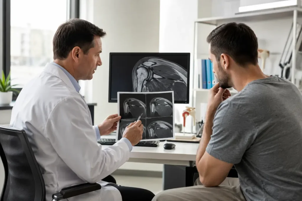
MRI is not a final diagnosis by itself. After the examination, the results should be shown to the doctor who referred you for diagnostics: an orthopedist, traumatologist, sports physician, rheumatologist, surgeon or another relevant specialist.
When is MRI of the shoulder joint prescribed?
MRI of the shoulder joint may be prescribed for pain, injury, arm weakness, limited movement, suspected tendon tear, shoulder instability or when other diagnostic methods have not provided enough information.
Shoulder MRI may be recommended if there is:
- shoulder pain during movement;
- pain when raising the arm;
- pain after a fall or impact;
- pain after training or physical work;
- limited shoulder movement;
- inability to move the arm behind the back;
- arm weakness;
- clicking or a feeling of instability in the shoulder;
- recurrent shoulder dislocations or subluxations;
- suspected tendon tear;
- suspected rotator cuff injury;
- suspected labral injury;
- shoulder pain that does not resolve after treatment;
- inflammatory process in the joint;
- follow-up after surgery;
- the need to clarify ultrasound, X-ray or CT results.
If after an injury the shoulder is deformed, the arm is severely limited in movement, pain is very intense, there is numbness, sensory disturbance or suspected dislocation or fracture, emergency medical care should be sought.
Shoulder MRI for pain
Shoulder pain may occur after injury, overload, training, physical work or gradually without an obvious cause. Patients often feel pain when raising the arm, moving the arm behind the back, getting dressed, sleeping on the side or performing usual everyday movements.
MRI of the shoulder joint may be useful if the pain:
- lasts a long time and does not resolve after treatment;
- worsens when raising the arm;
- interferes with sleeping on the side;
- appeared after a fall or impact;
- is accompanied by arm weakness;
- is combined with clicking or instability;
- occurs after sports or repetitive movements;
- is not explained by X-ray findings.
MRI helps the doctor understand whether the pain is related to tendons, the rotator cuff, labrum, bursa, capsule, cartilage, bone marrow edema or other changes inside the shoulder joint.
Shoulder MRI for rotator cuff injury
The rotator cuff — is a group of muscles and tendons that stabilize the shoulder joint and help move the arm. Rotator cuff injury — is one of the common causes of shoulder pain, especially in people over 40, athletes, manual workers and patients who often work with their arms overhead.
Rotator cuff problems may present as:
- pain when raising the arm;
- pain at night or while sleeping on the side;
- shoulder weakness;
- inability to raise the arm without pain;
- pain after exertion;
- limited movement;
- a shooting pain sensation in the shoulder.
MRI of the shoulder joint helps assess the rotator cuff tendons, detect inflammation, partial tear, complete tear, tendon retraction, bone marrow edema or associated changes in the joint. This is important for choosing treatment: physiotherapy, rehabilitation, injections, medication or surgery.
MRI of the shoulder joint after injury
After a shoulder injury, MRI may be needed if pain does not resolve, movements remain limited or there is suspected damage to tendons, ligaments, the labrum or soft tissues. Injury may occur after falling on the arm, an impact, a sudden pull, sports load or dislocation.
Shoulder MRI after injury may help detect:
- tendon tear;
- rotator cuff injury;
- labral injury;
- consequences of dislocation;
- ligament injury;
- bone marrow edema;
- occult fracture;
- bursal inflammation;
- fluid in the joint;
- combined shoulder injuries.
If the shoulder hurts after an injury, the arm does not lift or there is a feeling of instability, it is worth consulting a traumatologist or orthopedist and undergoing an examination as prescribed by the doctor.
Shoulder MRI for dislocation or instability
The shoulder joint has great mobility, so it may be prone to dislocations and recurrent instability. After dislocation, the labrum, capsule, ligaments, cartilage or bone structures may be damaged. If the shoulder “pops out”, there is a feeling of instability or recurrent subluxations, the doctor may refer the patient for MRI.
MRI of the shoulder joint can help assess:
- the labrum;
- the joint capsule;
- the ligament apparatus;
- consequences of previous dislocations;
- associated cartilage damage;
- bone marrow edema;
- soft tissue changes after injury.
In shoulder instability, it is important to correctly determine the extent of damage because treatment tactics depend on it: rehabilitation, load restriction, orthopedic treatment or surgery.
Shoulder MRI for sports injuries
The shoulder is often injured in sports: swimming, tennis, volleyball, basketball, martial arts, CrossFit, weight training, throwing sports and gymnastics. The cause of pain may be either an acute injury or gradual overload.
MRI of the shoulder joint after a sports injury may help detect:
- tendon tear or inflammation;
- rotator cuff injury;
- labral injury;
- biceps tendinopathy;
- bursitis;
- bone marrow edema;
- consequences of dislocation or subluxation;
- chronic soft tissue overload.
If shoulder pain interferes with training, recurs after exertion or limits arm movement, it is better not to continue exercising “through pain” and to consult a doctor.
MRI of the shoulder joint for osteoarthritis and inflammation
Shoulder osteoarthritis — is a degenerative process in which the articular cartilage, bone surfaces and periarticular tissues change. Inflammatory processes may affect the synovial membrane, tendons, bursae, capsule and other shoulder structures.
Shoulder MRI can help assess:
- the condition of articular cartilage;
- fluid in the joint;
- synovitis;
- bursitis;
- inflammatory changes in tendons;
- bone marrow edema;
- associated rotator cuff injuries;
- postoperative or scar changes.
To assess pronounced osteoarthritis, the doctor may also prescribe X-ray or CT, especially when bone deformities need to be seen. However, MRI provides more information about soft tissues, tendons, cartilage and inflammatory changes.
Shoulder MRI after surgery
After shoulder surgery, MRI may be needed to monitor the condition of the joint, assess postoperative changes, scar tissue, recurrent tendon or labral injury, as well as clarify the cause of pain, weakness or limited movement.
MRI after surgery may be prescribed if:
- pain persists after treatment;
- the shoulder remains weak;
- movements are limited;
- there is a recurrent feeling of instability;
- the condition after tendon repair needs to be assessed;
- postoperative changes need to be checked;
- the doctor is planning further treatment or rehabilitation.
If there are metal fixators or implants in the shoulder, you should report this when booking and bring documents related to the surgery if available.
MRI of the shoulder joint with and without contrast
In most cases, shoulder MRI is performed without contrast. This is often sufficient to assess tendons, the rotator cuff, ligaments, cartilage, the labrum, bursae, joint fluid, bone marrow edema, consequences of injury or degenerative changes.
MRI of the shoulder joint with contrast is not needed for everyone. Contrast enhancement may be prescribed when the doctor needs to clarify a tumor process, active inflammation, infectious involvement, postoperative changes or a complex clinical case where information without contrast may be insufficient.
You should not choose contrast on your own “for accuracy”. Contrast must be justified by the medical task. If the referral states MRI with contrast, you should report kidney diseases, allergic reactions, pregnancy, breastfeeding and medications you take when booking.
MRI of the shoulder joint price at HBMedical
At HBMedical, the cost of MRI of the shoulder joint depends on whether contrast enhancement is required.
| Service | Without contrast | With contrast |
| MRI of the shoulder joint / shoulder MRI | UAH 3400 | UAH 5660 |
The examination price includes the doctor’s report and a flash drive. It is better to clarify the current price of shoulder MRI when booking, because the final service depends on the doctor’s referral, the need for contrast and the specifics of the examination.
If you have a referral, tell the administrator its wording. This will help select the correct service: MRI of the shoulder joint without contrast, MRI with contrast or another diagnostic protocol.
How much does MRI of the shoulder joint cost in Kyiv?
At HBMedical, MRI of the shoulder joint costs from UAH 3400 without contrast. Shoulder MRI with contrast costs from UAH 5660. The price depends on the examination protocol and the need for contrast enhancement.
Contrast is not always required, so if the referral does not state “with contrast”, it is worth clarifying this with the doctor or when booking. You should not choose contrast enhancement on your own only because you want to “make it more accurate”.
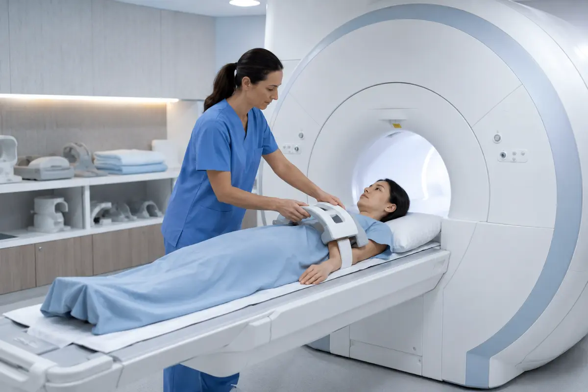
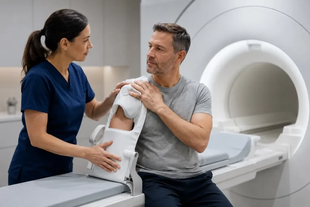
How is shoulder MRI performed?
The patient arrives at the medical center at the scheduled time, completes registration and fills out an MRI safety questionnaire. Medical staff check whether there are any contraindications to magnetic resonance imaging, implants or metal objects in the body.
Before the procedure, metal items, electronics, jewelry, watches, bank cards and other items that cannot be brought into the MRI room must be left outside. Then the patient lies down on the scanner table, and the shoulder is positioned to obtain clear images of the joint.
During scanning, it is important to remain still. Even small movements can reduce image quality. During operation, the machine makes loud sounds — this is a normal part of MRI. If the examination is performed with contrast, the contrast agent is administered intravenously through a catheter.
After the procedure, a radiologist analyzes the images and prepares a report. The results should be taken to the doctor who referred you for the examination.
How to prepare for MRI of the shoulder joint?
For shoulder MRI without contrast, no special complex preparation is usually required. The main thing is to inform the staff in advance about implants and metal structures and to bring previous examination results.
Before shoulder MRI, it is advisable to:
- bring your doctor’s referral if you have one;
- bring previous ultrasound, X-ray, CT, MRI, discharge summaries or laboratory test results;
- report metal structures, implants, clips or foreign metal bodies;
- report pregnancy or possible pregnancy;
- wear comfortable clothing without metal elements;
- remove jewelry, piercings, watch and metal accessories;
- do not bring a phone, bank cards, keys or electronic devices into the MRI room;
- clarify when booking whether additional preparation is needed in your specific case.
If MRI with contrast is prescribed, kidney function may need to be checked, in particular with a blood creatinine test. You should also report allergic reactions, kidney diseases, pregnancy or breastfeeding.
Can shoulder MRI be performed with metal or after surgery?
The possibility of MRI depends on the type of metal structure, its location and MRI compatibility. Many modern orthopedic structures are not an absolute contraindication, but they must be reported before the examination.
Before booking, tell the administrator if you have:
- screws, plates or fixators after surgery;
- an endoprosthesis or implant;
- metal fragments;
- a pacemaker or another electronic device;
- vascular clips;
- a cochlear implant;
- documents for the installed structure.
It is advisable to bring a postoperative discharge summary or implant passport if available. This will help medical staff assess the safety of performing MRI.
Are there contraindications to MRI of the shoulder joint?
MRI has limitations due to the strong magnetic field. Before the procedure, it is essential to report all implants, surgeries and foreign metal objects in the body.
MRI may be contraindicated or require additional approval in case of:
- a pacemaker or another electronic implant if it is not MRI-compatible;
- cochlear implants;
- some vascular clips;
- metal fragments or foreign bodies;
- certain types of prostheses or implants;
- the first trimester of pregnancy — by the doctor’s decision;
- inability to lie still;
- severe claustrophobia.
For MRI with contrast, it is also important to report severe kidney diseases, pregnancy, breastfeeding and previous reactions to contrast agents.
MRI, ultrasound, CT or shoulder X-ray — which should you choose?
X-ray, ultrasound, CT and MRI of the shoulder joint have different diagnostic capabilities. X-ray is more often used for the initial assessment of bones, fractures, dislocations, osteoarthritis and obvious bone changes.
Ultrasound can help assess the rotator cuff tendons, bursitis, fluid and some superficial soft tissues, but it is not always informative enough for deep intra-articular structures, the labrum or complex injuries.
CT better shows bone structures, complex fractures, post-traumatic changes and bone deformities. MRI is better suited for assessing tendons, the rotator cuff, ligaments, cartilage, the labrum, bone marrow, synovial membrane and hidden traumatic changes.
It cannot be said that one method is always better. The choice depends on symptoms, injury, the doctor’s referral and the diagnostic question.
Why have a shoulder MRI at HBMedical near Shuliavka?
HBMedical — is a modern medical center in Kyiv where you can undergo MRI of the shoulder joint on a Siemens Magnetom Sempra 1.5 Tesla scanner. The center is conveniently located near Shuliavska metro station, which is suitable for patients from Shevchenkivskyi, Solomianskyi, Sviatoshynskyi districts, KPI, Nyvky, Lukianivka, Dorohozhychi and other areas of Kyiv.
Advantages of HBMedical:
- convenient location near Shuliavska metro station;
- address: Kyiv, 3 O. Dovzhenka St.;
- modern Siemens Magnetom Sempra 1.5 Tesla MRI scanner;
- shoulder MRI with and without contrast;
- online or phone booking;
- diagnostics daily from 6:00 to 23:00;
- clear cost of MRI of the shoulder joint;
- the price includes the doctor’s report and a flash drive;
- possibility to undergo additional examinations or consultations in one medical center.
How to book a shoulder MRI in Kyiv?
To book a shoulder MRI at HBMedical, leave a request on the website or call the medical center. When booking, it is advisable to provide the exact wording from the referral: MRI of the shoulder joint, shoulder MRI with contrast, MRI of the right shoulder, MRI of the left shoulder or another protocol.
The administrator will help you choose a convenient time, clarify the current price, explain preparation and advise which documents or previous examination results you should bring with you.
HBMedical — shoulder MRI in Kyiv
Address: Kyiv, 3 O. Dovzhenka St., Shuliavska metro station
Services: shoulder MRI, MRI of the shoulder joint, shoulder MRI with contrast
Price: from UAH 3400
Booking: online or by phone
Frequently asked questions about shoulder MRI
Are shoulder MRI and MRI of the shoulder joint different examinations?
No, usually they are the same service. “Shoulder MRI” — is the shorter name patients often use. The more medically precise wording is “MRI of the shoulder joint”.
How much does MRI of the shoulder joint cost in Kyiv?
At HBMedical, MRI of the shoulder joint costs UAH 3400 without contrast and UAH 5660 with contrast. It is better to clarify the current price when booking.
What is the price of shoulder MRI without contrast?
MRI of the shoulder joint without contrast at HBMedical costs UAH 3400. The price includes the doctor’s report and a flash drive.
How much does MRI of the shoulder joint with contrast cost?
MRI of the shoulder joint with contrast at HBMedical costs UAH 5660. Contrast enhancement is performed according to indications or a doctor’s referral.
Where can I have MRI of the shoulder joint near Shuliavka?
MRI of the shoulder joint can be performed at the HBMedical medical center in Kyiv at the address: 3 O. Dovzhenka St., near Shuliavska metro station.
What does a shoulder MRI show?
Shoulder MRI shows the rotator cuff tendons, muscles, ligaments, cartilage, labrum, biceps tendon, bone marrow, joint fluid, inflammatory changes, consequences of injuries and postoperative changes.
When should MRI of the shoulder joint be performed?
MRI of the shoulder joint may be prescribed for pain, injury, limited movement, arm weakness, suspected tendon tear, rotator cuff injury, dislocation, instability, inflammation or postoperative changes.
Does MRI show shoulder tendon tears?
Yes, MRI helps assess the rotator cuff tendons and may show partial or complete tears, inflammation, degenerative changes and associated injuries.
Does MRI show labral injury of the shoulder?
MRI can help assess the labrum and changes related to shoulder instability or injury. In some complex cases, the doctor may recommend a special protocol or additional examination.
Is contrast needed for shoulder MRI?
In most cases, shoulder MRI is performed without contrast. Contrast is needed for specific indications, for example when a tumor, infectious, inflammatory or postoperative process is suspected.
Is preparation needed for MRI of the shoulder joint?
For shoulder MRI without contrast, no special complex preparation is usually required. You need to bring previous examination results, report implants or metal structures and come in clothing without metal elements.
Can shoulder MRI be performed after surgery?
The possibility depends on the type of surgery and the presence of metal structures. Before booking, you should report implants, screws, plates or other structures and bring medical documents if available.
Which is better: MRI or ultrasound of the shoulder joint?
Ultrasound may be useful for assessing superficial tendons and bursae, but MRI provides a more detailed image of tendons, ligaments, cartilage, the labrum, bone marrow and deep structures of the shoulder joint. The choice of method should be made by a doctor.
Is shoulder MRI painful?
No, the procedure itself is painless. The patient lies on the scanner table and must remain still. Discomfort may be related to shoulder pain, machine noise or the need for contrast administration.
Can I have a shoulder MRI without a referral?
It is advisable to have a doctor’s referral because it helps correctly determine the examination protocol, the need for contrast and the diagnostic area. If you do not have a referral, it is worth consulting an orthopedist, traumatologist, sports physician, rheumatologist or surgeon.


Саме до таких фахівців, в разі необхідності, є бажання повернутися!


Need specialist help?
Order a callback and our consultant will help you choose a doctor and make an appointment at a convenient time for you!